

Massive haemorrhage is common and often associated with high morbidity and mortality. We perform a systematic review of the literature, with extraction of the recommendations from the existing evidences because of the need for its improvement and the management standardization. From the results we found, we wrote a multidisciplinary consensus document. We begin with the agreement in the definitions of massive haemorrhage and massive transfusion, and we do structured recommendations on their general management (clinical assessment of bleeding, hypothermia management, fluid therapy, hypotensive resuscitation and damage control surgery), blood volume monitoring, blood products transfusion (red blood cells, fresh frozen plasma, platelets and their best transfusion ratio), and administration of hemostatic components (prothrombin complex, fibrinogen, factor VIIa, antifibrinolytic agents).
La hemorragia masiva es una entidad frecuente que se asocia a una elevada morbimortalidad. Ante la necesidad de la implementación y estandarización de su manejo, se realizó una revisión sistemática de la literatura, con extracción de recomendaciones en base a las evidencias existentes. A partir de las mismas se redactó un documento de consenso multidisciplinar. Desde las definiciones de hemorragia masiva y transfusión masiva, se establecen recomendaciones de actuación estructuradas en las medidas generales de manejo de las mismas (valoración clínica de la hemorragia, manejo de la hipotermia, reposición de la volemia, reanimación hipotensiva y cirugía de contención de daños), monitorización de la volemia, administración de hemocomponentes (concentrado de hematíes, plasma fresco, plaquetas, y óptima relación de administración entre ellos), y de hemostáticos (complejo protrombínico, fibrinógeno, factor VIIa, antifibrinolíticos).
Massive hemorrhage (MH) is a frequent condition caused by a range of circumstances including polytraumatism, peripartum, the perioperative period of different types of surgery, or gastrointestinal bleeding. It is commonly associated to important morbidity–mortality, conditioned to the underlying cause. In routine practice, the multidisciplinary management of MH is characterized by great variability; consensus-based recommendations therefore seem necessary in relation to the prevention, diagnosis, evaluation and application of the opportune therapeutic measures for controlling the bleeding.
One of the starting points is undoubtedly the need to establish an adequate definition of MH. The existing heterogeneity in interpreting the disorder means that it is difficult to establish the true incidence of MH in the different clinical scenarios; in this respect, it is considered that MH is often underestimated in terms of both its diagnosis and importance. Likewise, despite the different protocols that have been proposed to date, there is no agreement regarding the predictive value of the diagnostic tests, the best method for quantifying blood loss, the adequate assessment of treatment response, or the efficacy and safety of the recommended bleeding control measures.
Despite the efforts made in recent years to reach agreements regarding the protocols, guides and interventional algorithms referred to MH, and their diffusion, there is still an important lack of multidisciplinary consensus in our setting. The lack of scientific evidence in many of the proposed interventions, the undeniable urgency of the required treatment, and the lack of experience and specific training in the management of these situations found in some cases have pointed to the need for a consensus document, with the aim of facilitating the decision making process for all those involved in the treatment of MH. With this objective in mind, a series of experts belonging to the Spanish Society of Anesthesia and Resuscitation (Sociedad Española de Anestesiología y Reanimación, SEDAR), the Spanish Society of Intensive and Critical Care Medicine and Coronary Units (Sociedad Española de Medicina Intensiva, Crítica y Unidades Coronarias, SEMICYUC) and the Spanish Society of Thrombosis and Hemostasia (Sociedad Española de Trombosis y Hemostasia, SETH) decided to draft a manuscript with an eminently practical bearing, endorsed by the mentioned scientific societies. This consensus document offers the most important and appropriate recommendations in each case, extracted from the literature and applicable to MH in all its dimensions–from early diagnosis to the last step in management and control.
Form the multidisciplinary development of the document, we have underscored that its diffusion and application can contribute to improve the quality, safety and sustainability of the healthcare system, attempting to unify interventional and decision criteria.
As the authors of the initiative, we are convinced that the “Multidisciplinary consensus document on the management of massive hemorrhage (HEMOMAS document)” is an excellent tool with a permanent potential for modification and improvement, and that its usefulness will be confirmed over time, considering that Medicine is a very rapidly advancing field. The fundamental objective of our effort is to ensure that the document will be of help to all those involved in the management of MH. We hope to share our recommendations with these professionals, and to receive suggestions derived from their clinical practice.
MethodologyWith the scientific endorsement of the mentioned three scientific societies, a group of ten experts in anesthesia, intensive care and hematology was created, with the designation of a coordinator. These experts jointly developed the contents of the document. In the month of March 2013, a literature search was made of the Cochrane Library, Medline (PubMed) and Guiasalud databases, covering the last 10 years and using the combination of key words in the title or abstract: “massive hemorrhage”, “massive transfusion” and “hemorrhagic shock”. Based on the selected articles, we established the recommendations, categorizing the specific grades of recommendation and levels of evidence found. The Grades of Recommendation Assessment, Development and Evaluation (GRADE)1 were used to this effect, transferring the results to an alphanumerical scale in order to improve understanding of the recommendations2 (see Supplementary material online).
Results and discussionLiterature search and development of recommendationsThe literature search, carried out in March 2013, yielded a total of 4264 articles. After defining their priority through successive reading and analysis of the title, abstract and full text articles, we selected a total of 190 publications, to which 31 further articles (recently published or identified from the literature during the document drafting process) were added.
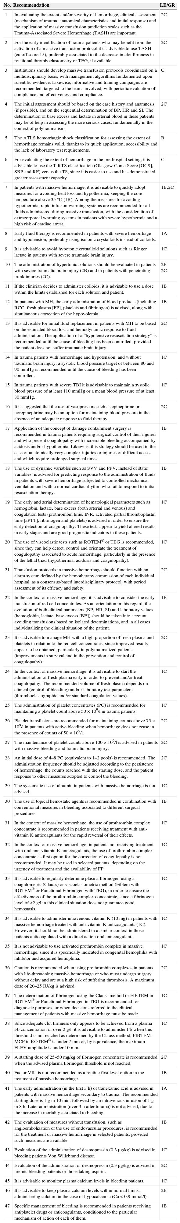
Following the literature synthesis and after application of the consensus methodology, we validated a total of 47 recommendations (Table 1).
Summary of the recommendations.
| No. | Recommendation | LE/GR |
|---|---|---|
| 1 | In evaluating the extent and/or severity of hemorrhage, clinical assessment (mechanism of trauma, anatomical characteristics and initial response) and the application of massive transfusion prediction scales such as the Trauma-Associated Severe Hemorrhage (TASH) are important. | 2C |
| 2 | For the early identification of trauma patients who may benefit from the activation of a massive transfusion protocol it is advisable to use TASH (cutoff score 15), preferably associated to the decrease in clot firmness in rotational thromboelastometry or TEG, if available. | 2C |
| 3 | Institutions should develop massive transfusion protocols coordinated on a multidisciplinary basis, with management algorithms fundamented upon scientific evidence. Likewise, informative and training campaigns are recommended, targeted to the teams involved, with periodic evaluation of compliance and effectiveness and compliance. | C |
| 4 | The initial assessment should be based on the case history and anamnesis (if possible), and on the sequential determination of BP, HR and SI. The determination of base excess and lactate in arterial blood in these patients may be of help in assessing the more serious cases, fundamentally in the context of polytraumatism. | 2C |
| 5 | The ATLS hemorrhagic shock classification for assessing the extent of hemorrhage remains valid, thanks to its quick application, accessibility and the lack of laboratory test requirements. | B |
| 6 | For evaluating the extent of hemorrhage in the pre-hospital setting, it is advisable to use the T-RTS classification (Glasgow Coma Score [GCS], SBP and RF) versus the TS, since it is easier to use and has demonstrated greater assessment capacity. | C |
| 7 | In patients with massive hemorrhage, it is advisable to quickly adopt measures for avoiding heat loss and hypothermia, keeping the core temperature above 35°C (1B). Among the measures for avoiding hypothermia, rapid infusion warming systems are recommended for all fluids administered during massive transfusion, with the consideration of extracorporeal warming systems in patients with severe hypothermia and a high risk of cardiac arrest. | 1B,2C |
| 8 | Early fluid therapy is recommended in patients with severe hemorrhage and hypotension, preferably using isotonic crystalloids instead of colloids. | 1A |
| 9 | It is advisable to avoid hypotonic crystalloid solutions such as Ringer lactate in patients with severe traumatic brain injury. | 1C |
| 10 | The administration of hypertonic solutions should be evaluated in patients with severe traumatic brain injury (2B) and in patients with penetrating trunk injuries (2C). | 2B-2C |
| 11 | If the clinician decides to administer colloids, it is advisable to use a dose within the limits established for each solution and patient. | 1B |
| 12 | In patients with MH, the early administration of blood products (including RCC, fresh plasma [FP], platelets and fibrinogen) is advised, along with simultaneous correction of the hypovolemia. | 1B |
| 13 | It is advisable for initial fluid replacement in patients with MH to be based on the estimated blood loss and hemodynamic response to fluid administration. The application of a “hypotensive resuscitation strategy” is recommended until the cause of bleeding has been controlled, provided the patient does not suffer traumatic brain injury. | 2C |
| 14 | In trauma patients with hemorrhage and hypotension, and without traumatic brain injury, a systolic blood pressure target of between 80 and 90mmHg is recommended until the cause of bleeding has been controlled. | 1C |
| 15 | In trauma patients with severe TBI it is advisable to maintain a systolic blood pressure of at least 110mmHg or a mean blood pressure of at least 80mmHg. | 1C |
| 16 | It is suggested that the use of vasopressors such as epinephrine or norepinephrine may be an option for maintaining blood pressure in the absence of an adequate response to fluid therapy. | 2C |
| 17 | Application of the concept of damage containment surgery is recommended in trauma patients requiring surgical control of their injuries and who present coagulopathy with incoercible bleeding accompanied by acidosis and/or hypothermia. Likewise, this strategy should be used in the case of anatomically very complex injuries or injuries of difficult access and which require prolonged surgical times. | 1B |
| 18 | The use of dynamic variables such as SVV and PPV, instead of static variables, is advised for predicting response to the administration of fluids in patients with severe hemorrhage subjected to controlled mechanical ventilation and with a normal cardiac rhythm who fail to respond to initial resuscitation therapy. | 1B |
| 19 | The early and serial determination of hematological parameters such as hemoglobin, lactate, base excess (both arterial and venous) and coagulation tests (prothrombin time, INR, activated partial thromboplastin time [aPTT], fibrinogen and platelets) is advised in order to ensure the early detection of coagulopathy. These tests appear to yield altered results in early stages and are good prognostic indicators in these patients. | 1C |
| 20 | The use of viscoelastic tests such as ROTEM® or TEG is recommended, since they can help detect, control and orientate the treatment of coagulopathy associated to acute hemorrhage, particularly in the presence of the lethal triad (hypothermia, acidosis and coagulopathy). | 1C |
| 21 | Transfusion protocols in massive hemorrhage should function with an alarm system defined by the hemotherapy commission of each individual hospital, as a consensus-based interdisciplinary protocol, with period assessment of its efficacy and safety. | 2C |
| 22 | In the context of massive hemorrhage, it is advisable to consider the early transfusion of red cell concentrates. As an orientation in this regard, the evolution of both clinical parameters (BP, HR, SI) and laboratory values (hemoglobin, lactate, base excess [BE]) should be taken into account, avoiding transfusions based on isolated determinations, and in all cases individualizing the clinical situation of the patient. | 1B |
| 23 | It is advisable to manage MH with a high proportion of fresh plasma and platelets in relation to the red cell concentrates, since improved results appear to be obtained, particularly in polytraumatized patients (improvements in survival and in the prevention and control of coagulopathy). | 2C |
| 24 | In the context of massive hemorrhage, it is advisable to start the administration of fresh plasma early in order to prevent and/or treat coagulopathy. The recommended volume of fresh plasma depends on clinical (control of bleeding) and/or laboratory test parameters (thromboelastographic and/or standard coagulation values). | 1C |
| 25 | The administration of platelet concentrates (PC) is recommended for maintaining a platelet count above 50×109/l in trauma patients. | 1C |
| 26 | Platelet transfusions are recommended for maintaining counts above 75×109/l in patients with active bleeding when hemorrhage does not cease in the presence of counts of 50×109/l. | 2C |
| 27 | The maintenance of platelet counts above 100×109/l is advised in patients with massive bleeding and traumatic brain injury. | 2C |
| 28 | An initial dose of 4–8 PC (equivalent to 1–2 pools) is recommended. The administration frequency should be adjusted according to the persistence of hemorrhage, the counts reached with the starting dose, and the patient response to other measures adopted to control the bleeding. | 2C |
| 29 | The systematic use of albumin in patients with massive hemorrhage is not advised. | 1C |
| 30 | The use of topical hemostatic agents is recommended in combination with conventional measures in bleeding associated to different surgical procedures. | 1B |
| 31 | In the context of massive hemorrhage, the use of prothrombin complex concentrate is recommended in patients receiving treatment with anti-vitamin K anticoagulants for the rapid reversal of their effects. | 1C |
| 32 | In the context of massive hemorrhage, in patients not receiving treatment with oral anti-vitamin K anticoagulants, the use of prothrombin complex concentrate as first option for the correction of coagulopathy is not recommended. It may be used in selected patients, depending on the urgency of treatment and the availability of FP. | 1C |
| 33 | It is advisable to regularly determine plasma fibrinogen using a coagulometric (Clauss) or viscoelastometric method (Fibtem with ROTEM® or Functional Fibrinogen with TEG), in order to ensure the effectiveness of the prothrombin complex concentrate, since a fibrinogen level of <2g/l in this clinical situation does not guarantee good hemostasis. | 1C |
| 34 | It is advisable to administer intravenous vitamin K (10mg) in patients with massive hemorrhage treated with anti-vitamin K anticoagulants (1C). However, it should not be administered in a similar context in those patients anticoagulated with a direct action oral anticoagulant. | 1C |
| 35 | It is not advisable to use activated prothrombin complex in massive hemorrhage, since it is specifically indicated in congenital hemophilia with inhibitor and acquired hemophilia. | 1C |
| 36 | Caution is recommended when using prothrombin complexes in patients with life-threatening massive hemorrhage or who must undergo surgery without delay and are at a high risk of suffering thrombosis. A maximum dose of 20–25IU/kg is advised. | 2C |
| 37 | The determination of fibrinogen using the Clauss method or FIBTEM in ROTEM® or Functional Fibrinogen in TEG is recommended for diagnostic purposes, or when decisions referred to the clinical management of patients with massive hemorrhage must be made. | 1C |
| 38 | Since adequate clot firmness only appears to be achieved from a plasma Fb concentration of over 2g/l, it is advisable to administer Fb when this threshold is not reached as determined by the Clauss method, FIBTEM-MCF in ROTEM® is under 7mm or, by equivalence, the maximum FLEV amplitude is under 10mm. | 1C |
| 39 | A starting dose of 25–50mg/kg of fibrinogen concentrate is recommended when the advised plasma fibrinogen threshold is not reached. | 2C |
| 40 | Factor VIIa is not recommended as a routine first level option in the treatment of massive hemorrhage. | 1B |
| 41 | The early administration (in the first 3h) of tranexamic acid is advised in patients with massive hemorrhage secondary to trauma. The recommended starting dose is 1g in 10min, followed by an intravenous infusion of 1g in 8h. Later administration (over 3h after trauma) is not advised, due to the increase in mortality associated to bleeding. | 1A |
| 42 | The evaluation of measures without transfusion, such as angioembolization or the use of endovascular procedures, is recommended for the treatment of massive hemorrhage in selected patients, provided such measures are available. | 1B |
| 43 | Evaluation of the administration of desmopressin (0.3μg/kg) is advised in bleeding patients Von Willebrand disease. | 1C |
| 44 | Evaluation of the administration of desmopressin (0.3μg/kg) is advised in uremic bleeding patients or those taking aspirin. | 2C |
| 45 | It is advisable to monitor plasma calcium levels in bleeding patients. | 1C |
| 46 | It is advisable to keep plasma calcium levels within normal limits, administering calcium in the case of hypocalcemia (Ca<0.9mmol/l). | 2B |
| 47 | Specific management of bleeding is recommended in patients receiving antiplatelet drugs or anticoagulants, conditioned to the particular mechanism of action of each of them. | 1B |
LE/GR: level of evidence/grade of recommendation.
The definition of MH is arbitrary and has little clinical value. However, any of the adequate definitions found in the literature can serve as a basis for implementing a specific MH management protocol. The most common definitions include the following:
- •
Blood loss of over 150ml/min during more than 10min.3
- •
Loss of a blood volume in 24h.
- •
Major hemorrhage requiring the transfusion of 4 red cell concentrate units in 1h.
- •
Loss of 1–1.5 volemia equivalents in 24h.4
- •
Loss of 50% of volemia equivalent in 3h.5
- •
Life-threatening major hemorrhage requiring massive transfusion.6
Although all of these definitions were considered adequate and applicable to routine practice, the panel of experts agreed that the first definition is probably the best when the blood losses are quantifiable–though such quantification is not always possible.
What is the definition of massive transfusion?In adults, massive transfusion (MT) can be defined as the transfusion of half the body blood volume in 4h, or of more than one such volume in 24h (the blood volume of an adult is about 70ml/kg).6
For quantification and comparison purposes, the most widely accepted definition is the administration of at least 10 red cell concentrate units in the 24h following the start of treatment.7,8
What are the most frequent causes of massive hemorrhage?No strict order in the frequency of causes of MH can be established, since it is largely dependent upon the clinical and social context involved. Nevertheless, in general it is agreed that the leading causes of MH are:
- •
Polytraumatism
- •
Cardiovascular surgery
- •
Postpartum hemorrhage
- •
Gastrointestinal bleeding
- •
Hepatobiliary surgery
It is necessary to identify patients who may suffer MH, based on the use of predictive scales. Different studies, mainly in the context of polytrauma patients, have developed and validated a number of systems and algorithms9–13 that facilitate such identification, particularly in the early stages of bleeding. In general, the more sophisticated systems, involving a larger number of variables, are better than the simpler designs–though prospective evaluations are still needed in order to improve the prediction process and further develop the existing scales.
Recommendation 19,13–22. In evaluating the extent and/or severity of hemorrhage, clinical assessment (mechanism of trauma, anatomical characteristics and initial response) and the application of massive transfusion prediction scales such as the Trauma-Associated Severe Hemorrhage (TASH) are important (2C).
Massive transfusionWhat clinical and/or biological parameters should activate the intervention protocol in the event of massive hemorrhage?The rapid identification of patients at risk of suffering MH and therefore of requiring MT is essential for immediate activation of the corresponding massive transfusion protocols (MTPs).23 A number of clinical scales and laboratory test parameters have been developed to objectively identify such patients.
Regarding the clinical scales, the TASH score13 offers the greatest sensitivity (84.4%) and specificity23 (78.4%). Another scale that is very widely used, since it does not involve laboratory test data that may delay the decision to activate the MTP, is the Assessment of Blood Consumption (ABC)24 (see Supplementary material online).
Compatible blood transfusion of patients with severe trauma upon admission to the emergency service has been shown to be an independent predictor of the need for early MT (>10 red cell concentrate units [RCC] in 6h), not only of blood but also of plasma and platelets.25
Regarding the laboratory test parameters upon arrival in hospital, a base deficit of >6mEq/l, plasma hemoglobin<11g/dl and pH<7.25 have been shown to significantly increase the risk of MT, and are included in several of the predictive scales.
In the study published by Davenport et al.26, involving 300 trauma patients, rotational thromboelastometry was used to identify acute coagulopathy and to predict the need for MT. A clot size after 5min of ≤35mm detected 71% of the patients who required MT, versus 43% who were detected by a prothrombin time of >1.2–with the added advantage that identification is established very quickly.
Other authors have reported similar results with both rotational thromboelastometry27 and thromboelastography (TEG).28
Recommendation 214. For the early identification of trauma patients who may benefit from the activation of a massive transfusion protocol it is advisable to use TASH (cutoff score 15), preferably associated to the decrease in clot firmness in rotational thromboelastometry or TEG, if available (2C).
Have the massive transfusion protocols been shown to improve patient survival?The use of a MTP has been shown to reduce both patient mortality and the need for blood product (BP) transfusions.29–31 The mechanisms underlying such improvement appear to be the administration of plasma and platelets in large proportions in relation to the red cell concentrates (RCC) in patients at high risk of requiring MT, as well as promptness in the start of transfusion,32 thereby allowing early management of the coagulopathy, earlier bleeding arrest, and a reduction of transfused blood products.
Borgman et al.32, in a multicenter retrospective study, showed the use of high plasma/RCC proportions (>1:2) in trauma patients at high risk of requiring MT (TASH score≥15) to be significantly correlated to improved survival (OR: 2.5 [1.6–4]). However, in patients at a lesser risk of requiring MT (TASH score<15), the use of such high proportions did not improve survival and significantly increased the risk of multiorgan failure (47% vs 38%).
Riskin et al.33, on evaluating mortality two years before and after the adoption of an MTP in trauma patients, recorded a significant decrease in mortality (45% vs 19%) after introduction of the protocol. In this study there were no significant alterations in either the proportionality or the total transfused volume of the blood products used in both groups. The main difference, to which the decrease in mortality after introduction of the MTP was attributed, was the promptness of the start of transfusion (the minutes taken to start transfusion of the specific blood group), with decreases of 39%, 33% and 42% for RCC, plasma and platelets, respectively.
Although the improvements in patient survival observed by most groups after the introduction of an MTP have been evidenced particularly in polytraumatized patients, other massive bleeding scenarios in which coagulopathy is suspected and bleeding control proves difficult can also derive benefit. However, no studies have produced sufficient evidence as to which protocol is best for improving patient survival.
On the other hand, before drawing conclusions on the efficacy of MTPs in reference to patient prognosis, due consideration is required of the analysis of both the protocol itself and of the degree of compliance.34,35
What criteria should be used to construct, implement and monitor a massive transfusion protocol?Massive transfusion protocols must offer a coordinated and efficient decision making process capable of ensuring the optimum management of MT.36 Enticott et al.37, in a systematic review, summarized the points considered crucial for guiding the design and implementation of an MTP. The steps considered necessary for such implementation are described in the supplementary material online.29–38
Recommendation 337. Institutions should develop massive transfusion protocols coordinated on a multidisciplinary basis, with management algorithms fundamented upon scientific evidence. Likewise, informative and training campaigns are recommended, targeted to the teams involved, with periodic evaluation of compliance and effectiveness and compliance (C).
General interventionsClinical assessment of hemorrhageWhat are the essential aspects in the initial clinical assessment of hemorrhage?In trauma patients we should sequentially evaluate systolic blood pressure (SBP), heart rate (HR) and respiratory frequency (RF), and calculate the shock index (SI=HR/SBP) recorded at the site of the accident and at the time of patient arrival in hospital. The differences between the measurements, referred to as ΔSBP and ΔHR, offer good prognostic discrimination capacity, though ΔSI is superior in predicting mortality after 48h in patients with moderate injuries, and is therefore of greater help in deciding treatment.39–42
The Triage Revised Trauma Score (T-RTS), developed for the in-ambulance identification of those trauma patients who may benefit from admission to a specialized hospital, does not require summing of the coded values, is easier to apply than the Trauma Score (TS), and has been shown to be substantially more useful from the prognostic perspective than the TS.
Recommendation 443–45. The initial assessment should be based on the case history and anamnesis (if possible), and on the sequential determination of BP, HR and SI. The determination of base excess and lactate in arterial blood in these patients may be of help in assessing the more serious cases, fundamentally in the context of polytraumatism (2C).
How should the extent of hemorrhage be evaluated?Although the Advanced Trauma Life Support (ATLS) scale was first described over 20 years ago by the American College of Surgeons for estimating blood loss and defining treatment–including the need or not for immediate surgical control–it remains a very useful tool. Depending on the blood volume lost, the ATLS is classified from grade I to grade IV according to the observed clinical consequences.22 This classification system is useful for stratifying the early manifestations and physiopathological signs related to the loss of blood (see Supplementary material online).
In addition to the above, increasingly frequent use is made of other scales for stratifying the severity of hemorrhage, such as the Triage Revised Trauma Score (T-RTS), which includes assessment of the Glasgow Coma Score (GCS), SBP and RF. In addition, the ROPE index (pulse rate over pressure evaluation) (HR divided by pulse pressure [PP, defined as the difference between SBP and the diastolic blood pressure (DBP)]; HR/PP) can be of help in detecting occult bleeding or patients at risk of suffering hemorrhagic shock. Different alternatives have been recommended for improving the discriminative capacity of the traditional vital signs (SBP, HR, RF and SI):
- •
The modified SI (MSI), corresponding to the ratio between HR and mean blood pressure (MBP), where MBP=([DBP×2]+SBP)/3.22
- •
The age-modified SI (ASI): ASI=SI×age.46
- •
The relation of HR to age, SBP/age (SBP/A), maximum HR for the age (MHR [mean heart rate]=220−age)−HR, and HR/MHR.47
The classification of hypovolemic shock based on the base deficit (BD) may be superior to the current ATLS for identifying the presence of hypovolemic shock, for adequately stratifying risk in bleeding patients, and for defining the need for blood product transfusions.48
Recommendation 539–41,49. The ATLS hemorrhagic shock classification for assessing the extent of hemorrhage remains valid, thanks to its quick application, accessibility and the lack of laboratory test requirements (B).
Recommendation 640,49–51. For evaluating the extent of hemorrhage in the pre-hospital setting, it is advisable to use the T-RTS classification (Glasgow Coma Score [GCS], SBP and RF) versus the TS, since it is easier to use and has demonstrated greater assessment capacity (C).
What is the lethal triad and how does it influence the outcome of these patients?The concurrence in patients with MH of hypothermia, acidosis and coagulopathy (lethal triad) worsens the prognosis.50,52,53 More recently, hypoxia and the hyperglycemia have been added as factors that also worsen the prognosis.54
Management of hypothermiaWhat is the temperature threshold beyond which the prognosis of patients with massive hemorrhage worsens?Hypothermia, defined as a core temperature of <35°C, is often observed in patients with hemorrhagic shock who require MT, and is associated to different complications such as:
- •
Diminished liver metabolism
- •
Lowered production of coagulation factors
- •
Platelet dysfunction
- •
Inhibition of the enzyme reactions of the coagulation cascade
Hypothermia is thus associated to increased bleeding, a greater need for transfusions, and increased mortality. In trauma patients, temperatures of under 35°C upon arrival in hospital are associated to an increased mortality risk,55 and this risk moreover increases as the temperature decreases further.56
In patients with MH it is therefore essential to adopt measures as quickly as possible for avoiding heat loss and for warming the patient (see Supplementary material). Although there is not enough evidence to recommend a concrete warming system in the context of MH, the data referred to fluid warming support the recommendation of warming all fluids administered during MT. In this regard, the systems based on countercurrent technology have been shown to be the most effective option for warming blood when the infusion rates are high.57 Extracorporeal warming systems should be considered in cases of severe hypothermia and a high risk of cardiac arrest, since they significantly shorten the time needed to warm the patient. Such systems are not without complications, however, particularly in relation to the vascular accesses.58
For each degree centigrade decrease in temperature there is a 10% decrease in coagulation factor activity59 and a 15% drop in the production of thromboxane B2, and thus in platelet aggregation.60 Below 33°C, the decrease in enzyme reactions of the coagulation cascade significantly blocks coagulation factor activity.61
Jurkovich et al.56, in a retrospective analysis of 71 adults with severe trauma, found that although the mortality rate for a temperature of 34°C or higher was 7%, this figure increased to 40% in the case of temperatures under 34°C, 69% for temperatures under 33°C, and 100% for temperatures under 32°C. In bleeding medical and surgical patients, temperatures of under 35°C are associated to increased blood loss.62
In a retrospective analysis of over 38,000 patients, Wang et al.55 found hypothermia at the time of admission to hospital in trauma patients to increase the mortality risk (OR: 4.04; 95% CI: 3.34–4.89); similar results have been recorded in patients with isolated brain injuries (OR; 3.14; 95% CI: 2.12–4.67).
Reynolds et al.63, in a prospective, multicenter cohort study of trauma patients with hemorrhagic shock, found the mean temperature in the first 24h after trauma to be under 34°C in 34% of the patients, and this temperature was moreover identified as an independent mortality risk factor in individuals receiving low plasma/red cell concentrate ratios, but not in those receiving high ratios.
Recommendation 755,56,59–64. In patients with massive hemorrhage, it is advisable to quickly adopt measures for avoiding heat loss and hypothermia, keeping the core temperature above 35°C (1B). Among the measures for avoiding hypothermia, rapid infusion warming systems are recommended for all fluids administered during massive transfusion, with the consideration of extracorporeal warming systems in patients with severe hypothermia and a high risk of cardiac arrest (2C).
Volume replacementWhat is the initial fluid of choice in patients with massive hemorrhage?In clinical practice, patients with severe hemorrhage and hypotension are administered intravenous fluids on an early basis. Preferably isotonic crystalloids are advised, avoiding hypotonic solutions such as Ringer lactate.
Among the isotonic crystalloids, balanced formulations in which the chloride levels are within the physiological limits (96–106mEq/l) is recommended.65
Recommendation 841. Early fluid therapy is recommended in patients with severe hemorrhage and hypotension, preferably using isotonic crystalloids instead of colloids (1A).
Recommendation 941. It is advisable to avoid hypotonic crystalloid solutions such as Ringer lactate in patients with severe traumatic brain injury (1C).
Recommendation 1041,65–69. The administration of hypertonic solutions should be evaluated in patients with severe traumatic brain injury (2B) and in patients with penetrating trunk injuries (2C).
What role do colloids play?The role of colloids in volume expansion in patients with MH is the subject of controversy and debate. The administration of certain colloids (hydroxyethyl starch [HES]) recently has been associated to kidney damage and to increased mortality in septic patients, though not in the context of administration as volume replacement strategy in hemorrhagic patients. In order to limit the adverse effects, if HES is used, it is advisable not to exceed a dose of 33–50ml/kg in 24h, depending on the type of starch, and renal function should be monitored during 90 days.67
Recommendation 1141,67. If the clinician decides to administer colloids, it is advisable to use a dose within the limits established for each solution and patient (1B).
At what point during massive hemorrhage should blood products be considered?The administration of blood products should be considered in the basic management of MH.
Recommendation 1241,70,71. In patients with MH, the early administration of blood products (including RCC, fresh plasma [FP], platelets and fibrinogen) is advised, along with simultaneous correction of the hypovolemia (1B).
What are the criteria and/or parameters guiding volume replacement?In addition to controlling the cause of hemorrhage, most studies recommend moderately restricted volume support, adopting the concept of hypotensive resuscitation if necessary, except in cases of severe traumatic brain injury.
Recommendation 1341,72. It is advisable for initial fluid replacement in patients with MH to be based on the estimated blood loss and hemodynamic response to fluid administration. The application of a “hypotensive resuscitation strategy” is recommended until the cause of bleeding has been controlled, provided the patient does not suffer traumatic brain injury (2C).
Hypotensive resuscitationSituations for the application of hypotensive resuscitationVolume replacement and BP restoration traditionally have been considered essential objectives in the management of trauma patients with hemorrhagic shock, and massive volume administration is needed in this respect. The normalization of BP without control of the bleeding source may further increase bleeding and therefore also result in greater fluid therapy needs–with an increased risk of coagulopathy, hypothermia and other complications which in turn contribute to worsen bleeding. This vicious circle can worsen the patient prognosis.
In recent years, and in the absence of traumatic brain injury (TBI), this strategy has shifted toward more conservative positions (so-called “hypotensive resuscitation”) that aim to ensure critical perfusion of vital organs for a short period of time, without affecting their function, and until the cause of bleeding has been identified and controlled. In penetrating trauma, the adoption of a conservative fluid therapy strategy has been shown to be effective,73 and although there is less supporting evidence in the case of closed trauma, a number of studies suggest that it may also be useful in this scenario.72,74–76
It is currently considered that in patients without TBI, the application of hypotensive resuscitation is advisable in situations of trauma with hemorrhagic shock until the source of bleeding has been controlled.41
Recommendation 1441,72–76. In trauma patients with hemorrhage and hypotension, and without traumatic brain injury, a systolic blood pressure target of between 80 and 90mmHg is recommended until the cause of bleeding has been controlled (1C).
In patients with TBI, the presence of hypotension is associated to increased mortality and a poorer functional outcome.77,78 Therefore, in cases of severe TBI, it is advisable to maintain a SBP of at least 110mmHg, or a MBP of at least 80mmHg.41
Recommendation 1541,77,78. In trauma patients with severe TBI it is advisable to maintain a systolic blood pressure of at least 110mmHg or a mean blood pressure of at least 80mmHg (1C).
When should vasopressors be used in the context of massive hemorrhage?In some cases, volume replacement alone is not enough to correct the hemodynamic situation. In such cases the risk of hypoperfusion of vital organs is high, and the use of vasoactive drugs can be decided to help revert the situation. The use of vasopressors is not without controversy, however, and one study has suggested that their administration in the first 12h is associated to a mortality increase of almost 80%.79 Nevertheless, this study has been criticized due to methodological reasons, and it is presently considered that if the patient fails to respond to fluid therapy, vasopressors can be used to restore the perfusion of vital organs.41
Recommendation 1641,79. It is suggested that the use of vasopressors such as epinephrine or norepinephrine may be an option for maintaining blood pressure in the absence of an adequate response to fluid therapy (2C).
Damage containment surgeryUnder what circumstances should damage containment surgery be considered?The concept of damage containment surgery refers to surgery of short duration, designed to control situations that cannot be postponed in critical patients with scant physiological reserves. These patients often have hemodynamic problems and uncontrolled bleeding, with coagulation disorders and perfusion alterations with metabolic acidosis and/or hypothermia. Such cases generally involve abdominopelvic injuries, though the same concept can be extended to patients with similar characteristics involving other types of injuries requiring urgent surgery. After emergency control has been achieved, the patient is stabilized in the Intensive Care Unit (ICU), and definitive surgical treatment is provided at a later stage. A recent Cochrane review80 acknowledges that no randomized studies have analyzed the efficacy of this strategy, though a series of retrospective studies confirm its usefulness,81–83 and the indication of damage containment surgery is currently contemplated by some treatment guides.41
Recommendation 1741,80–83. Application of the concept of damage containment surgery is recommended in trauma patients requiring surgical control of their injuries and who present coagulopathy with incoercible bleeding accompanied by acidosis and/or hypothermia. Likewise, this strategy should be used in the case of anatomically very complex injuries or injuries of difficult access and which require prolonged surgical times (1B).
MonitoringVolume monitoringWhat is the best method for the continuous monitoring of patient response to volume expansion?Volume expansion is the first measure in the management of MH. However, the excess of fluids can have harmful effects.84 Although the determination of intravascular volume is one of the most difficult challenges in clinical practice, it is essential to optimize this volume through reliable predictors of patient response to fluids. A number of static and dynamic hemodynamic variables are available for this purpose.
It has been shown that central venous pressure is of little predictive value in determining the hemodynamic response to volume loading.85,86 Other investigated static variables such as pulmonary capillary pressure or global end-diastolic volume likewise have not demonstrated greater predictive efficacy.87
The gold standard for monitoring the response to fluid loading is the continuous monitoring of cardiac output.88 The absence of an increase in cardiac output in response to volume loading indicates that the plateau of the cardiovascular function curve has been reached. This in turn advises great caution in the administration of fluids, in order to prevent venous congestion.89
In place of static hemodynamic variables, the recommendation is to use dynamic indicators which allow us to assess preload with a view to predicting the response to fluids in patients with controlled mechanical ventilation and a normal cardiac rhythm–though in patients with severe hypovolemia due to evident bleeding, initial fluid loading to assess the response is not necessary.90 The most widely used dynamic variables and with the greatest predictive value have been found to be stroke (systolic) volume variation (SVV) and pulse pressure variation (PPV).91–94 Starting the sequential evaluation of cardiac function with echocardiography in shock patients, before resorting to more invasive techniques, has also been suggested.90
Recommendation 1894,95. The use of dynamic variables such as SVV and PPV, instead of static variables, is advised for predicting response to the administration of fluids in patients with severe hemorrhage subjected to controlled mechanical ventilation and with a normal cardiac rhythm who fail to respond to initial resuscitation therapy (1B).
Laboratory monitoringWhat laboratory test parameters are most useful for the management of massive hemorrhage?Among the laboratory test parameters of greatest usefulness in assessing the evolution of patients with MH, mention must be made of the early and serial determination of basic hematological values such as hemoglobin, lactate, base excess (BE) and coagulation parameters (prothrombin time [PT], activated partial thromboplastin time [aPTT], fibrinogen1 and platelets) for detecting the presence of coagulopathy is soon as possible–particularly in the presence of hypothermia and acidosis. These tests appear to yield altered results in early stages and are good prognostic indicators in these patients. The inclusion of arterial lactate, in addition to base excess, contributes to differentiate the most seriously ill patients.
The adoption of so-called point of care devices (used to monitor blood parameters at the patient bedside) has greatly improved the availability and utilization of these tests in evaluating patients with MH. They have also shortened the time required for the sample to reach the place where it is processed, and the time required to obtain the results.
Furthermore, the utilization of viscoelastic tests such as thromboelastography (TEG) and rotational thromboelastometry (ROTEM®) can help detect, control and orientate the treatment of coagulopathy associated to acute hemorrhage, particularly in the presence of the lethal triad (hypothermia, acidosis and coagulopathy).
Among the laboratory data used to monitor coagulopathy, mention must be made of the early and serial determination of aPTT, PT, the International Normalized Ratio (INR), fibrinogen (Fb) via the Clauss method, and platelets.41 Likewise, in surgical or trauma patients with severe hemorrhage, completion of the coagulation study with TEG or ROTEM® should be considered, conditioned to their availability in each center.96,97
Recommendation 1941,43,44,98. The early and serial determination of hematological parameters such as hemoglobin, lactate, base excess (both arterial and venous) and coagulation tests (prothrombin time, INR, activated partial thromboplastin time [aPTT], fibrinogen and platelets) is advised in order to ensure the early detection of coagulopathy. These tests appear to yield altered results in early stages and are good prognostic indicators in these patients. (1C).
Recommendation 2099,100. The use of viscoelastic tests such as ROTEM®or TEG is recommended, since they can help detect, control and orientate the treatment of coagulopathy associated to acute hemorrhage, particularly in the presence of the lethal triad (hypothermia, acidosis and coagulopathy) (1C).
Administration of blood componentsRed cell concentrateWhat are the criteria for the transfusion of red cell concentrates in a patient with massive hemorrhage?In general, terms, both the European and the Australian guides suggest the avoidance of red cell concentrate transfusions in the presence of hemoglobin values above 10g/dl,60,93 though special consideration is indicated in the case of low-weight patients and elderly individuals.101
In certain scenarios such as heart surgery and patients with ischemic heart disease, hemoglobin levels of above 9g/dl should be reached.102,103 However, in patients who are clinically stable, more restricted transfusion criteria apply, with hemoglobin levels of 8g/dl.104
Recommendation 216,93,101–104. Transfusion protocols in massive hemorrhage should function with an alarm system defined by the hemotherapy commission of each individual hospital, as a consensus-based interdisciplinary protocol, with period assessment of its efficacy and safety (2C).
Recommendation 226,93,105. In the context of massive hemorrhage, it is advisable to consider the early transfusion of red cell concentrates. As an orientation in this regard, the evolution of both clinical parameters (BP, HR, SI) and laboratory values (hemoglobin, lactate, base excess [BE]) should be taken into account, avoiding transfusions based on isolated determinations, and in all cases individualizing the clinical situation of the patient (1B).
Optimum treatment ratiosIs there an optimum ratio for the administration of blood components in the management of massive hemorrhage?There is great controversy regarding the optimum ratio for the administration of blood components in the management of massive hemorrhage. Initial studies suggested that a FP:RCC ratio of 1:1 reduces mortality in war injuries.106,107 Additional cohort studies appeared to indicate a beneficial effect of the 1:1 ratio in other massive bleeding scenarios. In 2010, however, a more detailed review of the United States Army in 777 trauma patients transfused with the 1:1 ratio was unable to demonstrate a decrease in mortality.108 In contrast, different studies pointed to the possibility that 1:1 protocols might induce undesirable effects that outweigh the benefits.109–112 It was found that:
- 1)
The use of fixed-ratio protocols caused patients who do not need plasma to receive it needlessly.
- 2)
The plasma in excess received by the patients generated problems of fluid overload, increased the risk of multiorgan failure, and raised the number of complications (respiratory distress, multiorgan failure and infections).
There is no evidence that transfusion strategies based on fixed-ratio protocols, with the administration of red cell concentrates/fresh plasma/platelet concentrates (RCC:FP:PC) in 1:1:1 proportion, offer a favorable risk-benefit relationship in MH. Furthermore, there is a high risk that the use of strategies of this kind in non-trauma patients may result in more undesirable effects than benefits.113 In sum, although benefits have been suggested with the use of fixed-proportion (1:1:1 or 2:1:1) transfusion strategies, it has not been possible to demonstrate the superiority of one strategy over the rest. In some situations, the use of fixed-ratio protocols can cause complications. A more detailed study is required of the benefit-risk profile of the 1:1:1 transfusion protocol in patients with severe trauma.
Recommendation 236,41,93. It is advisable to manage MH with a high proportion of fresh plasma and platelets in relation to the red cell concentrates, since improved results appear to be obtained, particularly in polytraumatized patients (improvements in survival and in the prevention and control of coagulopathy) (2C).
Fresh plasmaHow and when should fresh plasma be used?The transfusion of FP remains the standard for preventing and treating coagulopathy in MH, though its inconveniences include the risk of transfusion-associated circulatory overload (TACO) (more frequent in the presence of heart failure), transfusion-related acute lung injury (TRALI), or the need in some cases to wait for as long as 45min for transfusion to become available.114
Recommendation 24115–118. In the context of massive hemorrhage, it is advisable to start the administration of fresh plasma early in order to prevent and/or treat coagulopathy. The recommended volume of fresh plasma depends on clinical (control of bleeding) and/or laboratory test parameters (thromboelastographic and/or standard coagulation values) (1C).
PlateletsWhat is the limiting circulating platelet count for starting the transfusion of platelet concentrates?There is no solid scientific evidence regarding the platelet count capable of guaranteeing hemostasis. In general, use is made of the opinions and/or conclusions of previous studies.41 The base concept is that in normal situations the transfusion of platelets is not necessary in patients with counts of over 100×109/l. The optimum count for restoring hemostasis in actively bleeding patients is not known.
There is some agreement that the platelet counts should be kept above 50×109/l in patients with active bleeding.119,120 Some experts advocate a higher platelet count (75×109/l) in patients with active bleeding or in which bleeding persists with counts of 50×109/l.121,122 Counts above 100×109/l would be more adequate for polytraumatized patients with brain injuries and massive bleeding.
No platelet count limits have been specifically defined for massive gynecological hemorrhage, liver transplantation or cardiovascular surgery, and when disseminated intravascular coagulation (DIC) with or without hyperfibrinolysis is moreover present, the indication of platelet administration must be based on the severity of bleeding and the specific conditions that may have given rise to MH. Therefore, given the platelet dysfunctions associated to these clinical situations, the limiting platelet count should not be used as reference.
Recommendation 2541,119–122. The administration of platelet concentrates (PC) is recommended for maintaining a platelet count above 50×109/l in trauma patients (1C).
Recommendation 2641,121–123. Platelet transfusions are recommended for maintaining counts above 75×109/l in patients with active bleeding when hemorrhage does not cease in the presence of counts of 50×109/l (2C).
Recommendation 2741,119,121,124. The maintenance of platelet counts above 100×109/l is advised in patients with massive bleeding and traumatic brain injury (2C).
What platelet dose should be administered, and with what frequency?There is no solid scientific evidence in this regard. The opinions come from publications of the Working Group created in 2005, and which has published guides in 2007, 2010 and 2013. Hospitals can receive platelets in the form of PC fractionated from whole blood units, pools obtained from mixing 4–5 concentrates (one pool being equivalent to 4–5 concentrates), or units obtained through single donor apheresis. There are no recommendations regarding the frequency of administration.
The increases in platelet counts are greater in the case of platelet transfusions obtained through identical ABO group apheresis, and also in the case of platelets stored for three days compared with 4–5 days.125 The origin (apheresis versus pool), ABO compatibility, and the duration of storage have a moderate effect upon the absolute and corrected post-transfusion platelet count increments, but have no measurable impact upon the prevention of the hemorrhage.125–128 In thrombocytopenic patients, PC not subjected to biological inactivation produce greater post-transfusion platelet count increments, but there is no evidence suggesting greater hemostatic efficacy in such cases.129 No studies have been made in patients with massive hemorrhage.
Recommendation 2841,100,119,122,126,130. An initial dose of 4–8 PC (equivalent to 1–2 pools) is recommended. The administration frequency should be adjusted according to the persistence of hemorrhage, the counts reached with the starting dose, and the patient response to other measures adopted to control the bleeding (2C).
What is the order of administration of platelet concentrates with respect to other blood products (plasma, red cell concentrates or other hemostatic agents)?An increase in hematocrit to 30% reduces the risk of hemorrhage in patients with thrombocytopenia. Liumbruno et al.122 recommend trying to raise hematocrit to close to 30% in order to reduce the risk of hemorrhage (recommendation 1C+). Increasing the hematocrit to 30% improves platelet hemostatic efficacy.131–134 In patients with MH, it seems to be advisable to transfuse PC after having administered red cell concentrates and FP.131
What are the side effects of the administration of platelets? What safety aspects should be taken into account to avoid them?The transfusion of platelets can produce adverse reactions of variable severity. One out of every 50,000 platelet units may be contaminated and cause complications secondary to severe sepsis. The storage and form of production of the platelets (pool, buffy coat or apheresis) can modify the biological response to the transfused concentrates and either reduce (apheresis) or increase (pool) patient exposure to one or more donors. The biological risk increases in patients subjected to massive transfusion, and the associated severity can increase depending on the biocomponent ratios used.135 Although there is no evidence of any clinical benefit from the use of different concentrates, storage times or treatments for reducing the presence of pathogens, the administration of concentrates stored for shorter periods of time, and the use of apheresis units, appears to reduce the biological risks.
The transfusion of FP and PC can cause transfusion-related acute lung injury (TRALI), which is infrequent and may go unnoticed, but is associated to high mortality rates. It is more common after the administration of FP, but has also been related to PC, which are vehiculized with plasma.136 The utilization of plasma from male blood donations, the use of lesser plasma volumes in the concentrates, and the utilization of single donors versus pools all contribute to reduce the risk of these complications.
Other blood componentsIs the use of albumin justified in patients with massive hemorrhage?The administration of albumin remains a controversial issue in the management of MH, since there are no conclusive studies supporting this practice. Albumin has not been found to be superior in volume replacement when compared with artificial colloids and crystalloids.137,138
Albumin formulations are available at concentrations of 5% and 20%. The lesser concentration solution (5%) is the only formulation indicated for volume replacement. The 20% concentration is exclusively administered for the treatment of severe hypoproteinemia requiring replacement therapy.139
Recommendation 29137–139. The systematic use of albumin in patients with massive hemorrhage is not advised (1C).
What is the role of topical hemostatic agents in the control of hemorrhage?The ideal topical hemostatic agent is that which affords rapid local bleeding control without affecting the systemic coagulation mechanisms. Furthermore, it should not be able to cross the filtering systems, and should not be of human or bovine origin, in order to avoid the transmission of infections. These characteristics can be offered by recombinant agents that contain chitosan, thereby obviating the risk of transmission of viral diseases.140,141 Topical agents have been successfully used in different surgical procedures, including heart, urological, gynecological and gastrointestinal surgery, and trauma surgery.
Recommendation 3041,140,141. The use of topical hemostatic agents is recommended in combination with conventional measures in bleeding associated to different surgical procedures (1B).
Administration of hemostatic agentsProthrombin complexWhat are the indications of prothrombin complex concentrate in the context of MH?In relation to the administration of prothrombin complex concentrate (PCC), the panel of experts recommends that both the indication and the dose and frequency of administration of PCC should be based on the specifications of the Summary of Product Characteristics.
In the case of patients anticoagulated with anti-vitamin K drugs (warfarin or acenocoumarol) in the context of MH, we can use PCC as an effective alternative to plasma for the urgent reversal of the anticoagulant effect.142 The starting dose of PCC (preferably comprising 4 factors, which is the formulation available in our setting) should be 50IU/kg,143 associated to a vitamin K dose of 10mg i.v. Since the thrombogenic side effects of PCC have been associated to high doses and successive dose administrations, it is advisable to evaluate the INR value before a new PCC dose is administered: if the INR<1.5, another dose of PCC is not advised–though the clinical parameters also must be taken into account.144
In patients with MH not associated to the use of anti-vitamin K anticoagulants, the use of PCC as first line treatment is generally not recommended.143,144 Prothrombin complex concentrate can be used in selected cases, fundamentally in situations where FP is not timely available, if the patient is at risk of suffering transfusion-associated circulatory overload (TACO), or in the presence of a risk of TRALI.
Recommendation 3193. In the context of massive hemorrhage, the use of prothrombin complex concentrate is recommended in patients receiving treatment with anti-vitamin K anticoagulants for the rapid reversal of their effects (1C).
Recommendation 3293. In the context of massive hemorrhage, in patients not receiving treatment with oral anti-vitamin K anticoagulants, the use of prothrombin complex concentrate as first option for the correction of coagulopathy is not recommended. It may be used in selected patients, depending on the urgency of treatment and the availability of FP (1C).
Recommendation 33105. It is advisable to regularly determine plasma fibrinogen using a coagulometric (Clauss) or viscoelastometric method (Fibtem with ROTEM®or Functional Fibrinogen with TEG), in order to ensure the effectiveness of the prothrombin complex concentrate, since a fibrinogen level of <2g/l in this clinical situation does not guarantee good hemostasis (1C).
Recommendation 34143. It is advisable to administer intravenous vitamin K (10mg) in patients with massive hemorrhage treated with anti-vitamin K anticoagulants (1C). However, it should not be administered in a similar context in those patients anticoagulated with a direct action oral anticoagulant (1C).
Is the use of activated prothrombin complex recommended in the context of massive hemorrhage?Recommendation 35145,146. It is not advisable to use activated prothrombin complex in massive hemorrhage, since such treatment is specifically indicated in congenital hemophilia with inhibitor and acquired hemophilia (1C).
What are the required safety precautions when using prothrombin complex?Prothrombin complex concentrate is generally safe, though some complications (fundamentally of a thrombotic nature) have been documented in certain scenarios.147
It is advisable to use PCC with caution in patients with thrombotic or cardiovascular risk factors, in individuals with artificial heart valves, with a history of ischemic stroke, or in patients subjected to revascularization after acute coronary syndrome, taking care not to exceed a dose of 20–25IU/kg. Likewise, PCC should be used with caution in polytraumatized patients, due to the risk of suffering thromboembolic events.143,147–149
Recommendation 36147,148. Caution is recommended when using prothrombin complexes in patients with life-threatening massive hemorrhage or who must undergo surgery without delay and are at a high risk of suffering thrombosis. A maximum dose of 20–25IU/kg is advised (2C).
FibrinogenDetermination of fibrinogenFibrinogen (Fb) is the factor exhibiting the earliest decrease in concentration after severe hemorrhage.150,151 The earliness and intensity of this decrease in concentration is predictive of the severity of hemorrhage.152
A number of techniques are available for the determination of Fb concentration, and their incorrect application or interpretation can complicate the diagnosis and decisions regarding the transfusion strategy to be used. The determinations of Fb derived from prothrombin time using automated coagulometers are not reliable for the management of MH. The Clauss technique is recommended for diagnostic purposes, or when decisions referred to the clinical management of patients with MH must be made.153 The determination of FIBTEM with ROTEM® or Functional Fibrinogen in TEG allows us to quickly detect alterations in the Fb levels in trauma patients. In this regard, it has been confirmed that the determination of Fb using the FIBTEM test in ROTEM® is closely correlated to the values obtained with the Clauss method.154,155 The Fb values may be erroneous in samples from hemodiluted patients, and particularly in those who have received colloids. The determination of Fb via FIBTEM in ROTEM® or of Functional Fibrinogen in TEG is a useful option and is faster than the determination of Fb using the Clauss method.
Recommendation 3799,153–155. The determination of fibrinogen using the Clauss method or FIBTEM in ROTEM®or Functional Fibrinogen in TEG is recommended for diagnostic purposes, or when decisions referred to the clinical management of patients with massive hemorrhage must be made (1C).
What is the plasma fibrinogen threshold for the administration of fibrinogen?Fibrinogen is the main coagulation substrate and the most abundant factor in plasma. However, it is also the factor that decreases fastest to critical levels in the case of MH.150
The bleeding tendency increases when the Fb concentration drops to below 1.5–2.0g/l.152,156,157 Recent European guides recommend the administration of Fb when the abovementioned threshold is not reached,41,93 considering that clot firmness increases linearly with the concentration of Fb, and that a minimum threshold of 2g/l is needed to reach optimum clot formation.158 However, further studies are needed to consolidate the evidence supporting this recommendation.159
On the other hand, rapid coagulation testing is essential for the early treatment of MH. An excellent correlation has been observed between the concentration of Fb and all the ROTEM® parameters, particularly FIBTEM.160 In effect, in both pediatric surgery and in polytraumatized patients,155,161 a maximum clot firmness (MCF) in FIBTEM of between 7 and 10mm is associated to plasma Fb levels of 1.5–2g/l. It is considered that FIBTEM-MCF <7mm indicates alteration of fibrin polymerization and a decrease in Fb concentration to under 1.5–1.8g/l.27,155,162,163 Calculation of the Fb levels via TEG indicates a normal range of Functional Fibrinogen Level (FLEV) values of between 2 and 4.45g/l, with a good correlation to the Clauss method.164
Recommendation 3841,93,150,152,155–158,160,161,163,165–167. Since adequate clot firmness only appears to be achieved from a plasma Fb concentration of over 2g/l, it is advisable to administer Fb when this threshold is not reached as determined by the Clauss method, FIBTEM-MCF in ROTEM®is under 7mm or, by equivalence, the maximum FLEV amplitude is under 10mm (1C).
What is the fibrinogen starting dose?The Fb concentrate dose should be individualized according to the severity of bleeding and the current plasma Fb concentration. The dose is calculated based on the following formula168:
Fb dose (g)=desired Fb increment (g/l)×plasma volume (l)
If we assume a plasma volume of 0.04l/kg, this formula shows that for each g of Fb administered, the plasma Fb level increases an average of 0.25–0.28g/l.169
However, when Fb replacement is guided by rotational thromboelastometry, the dose is calculated as follows:170
Fb dose (g)=FIBTEM-MCF (mm) target−actual FIBTEM-MCF (mm)× weight (kg)/140
On an approximate basis, according to this formula, for each g of Fb administered, FIBTEM-MCF increases 2mm.171 A similar calculation can be made for TEG based on the correlation of FLEV to the values of the Clauss method.164
Different studies have been published on the correction of coagulopathy or reduction of the transfusion requirements and the administration of Fb. As an example, in trauma patients, non-randomized studies have shown the administration of Fb to be effective in securing the mentioned objectives.99,172,173 Likewise, in orthopedic surgery, the administration of Fb appears to revert dilutional coagulopathy.174 In a randomized study of surgical patients subjected to radical cystectomy, the administration of Fb was found to reduce the transfusion needs.165 More studies have been published in recent years in relation to cardiovascular surgery, involving both randomized175 and non-randomized designs,170,175–179 and demonstrating that the administration of Fb reduces the transfusion needs. Many of these authors have used a model to individualize the Fb dose.175 Thus, when bleeding in 5min totals 60–250g, they administered a Fb dose calculated to reach a FIBTEM-MCF of approximately 22mm, equivalent to a plasma level of 3.6g/l, which would optimize the interaction between Fb and the available thrombin after cardiopulmonary bypass, and would improve hemostasis.176
Regarding the prophylactic use of Fb, a randomized coronary surgery study has been published in which the authors administered 2g of Fb to patients with a preoperative plasma level of under 3.8g/l, with confirmation of a decrease in postoperative bleeding.180 In contrast, another randomized multicenter study has not confirmed the efficacy of the preventive administration of 2g of Fb in postpartum hemorrhage among patients with normal blood fibrinogen levels.181
There are also two systematic reviews and a Cochrane review which have concluded that treatment with Fb reduces the transfusion needs and postoperative bleeding.159,182,183 However, in surgical patients it has been seen that the plasma Fb increments are short-lasting, and that 24h after surgery the Fb levels are similar to those recorded in the patients who received no such treatment.165,180,184
Controlled studies are still needed to investigate the possibility of thromboembolic events related to the administration of Fb.
In sum, more controlled studies are needed to evidence the efficacy of Fb and its use in the different clinical scenarioss.159 Likewise, it should be underscored that cryoprecipitates are no longer used as a source of Fb in many European countries, due to their adverse effects and difficult dosing characteristics, and have been replaced by Fb concentrates.185–189
Recommendation 3941,93,165. A starting dose of 25–50mg/kg of fibrinogen concentrate is recommended when the advised plasma fibrinogen threshold is not reached (2C).
Factor VIIaWhat are the indications for the administration of factor VIIa in the context of massive hemorrhage?The approved indications of factor VIIa are the prevention and treatment of hemorrhage in patients with congenital hemophilia A or B, congenital factor VII deficiency, acquired hemophilia and Glanzmann's thrombasthenia at a dose of 90μg/kg every 2h until the bleeding episode ceases.190–192
Regarding use outside its established indications, ex vivo studies and investigations in healthy volunteers have shown that factor rVIIa is able to antagonize the effect of many anticoagulants,193–195 including the new oral anticoagulants and vitamin K antagonists.196–198 However, its use is not advised for the routine reversal of coumarin-induced coagulopathy,199,200 since it is unable to reduce mortality when used for this purpose.201 In view of the lack of clinical evidence in favor of its effectiveness, and since some studies have reported an increase in thrombosis in patients administered factor rVIIa, its administration should only be considered as a last option in patients with bleeding complications who are receiving treatment with anticoagulants that lack a specific antidote or in which bleeding proves incoercible despite application of all the necessary measures.193,202–204 A single dose of 90μg/kg is recommended.205,206
Recommendation 40190–206. Factor VIIa is not recommended as a routine first level option in the treatment of massive hemorrhage (1B).
Antifibrinolytic agentsIn what context are antifibrinolytic agents used in massive hemorrhage?Hyperfibrinolysis is a basic component in different medical and surgical scenarios involving MH. This would justify the use of antifibrinolytic agents such as tranexamic acid (TXA) or ¿-aminocaproic acid (EACA). However, TXA has shown superiority over EACA in controlling bleeding, and there is little scientific evidence supporting the use of the latter drug.207,208 The results of the CRASH-2 study show the early administration of TXA to be indicated in patients with hemorrhage associated to trauma.209
Recommendation 4141,93,209,210. The early administration (in the first 3h) of tranexamic acid is advised in patients with massive hemorrhage secondary to trauma. The recommended starting dose is 1g in 10min, followed by an intravenous infusion of 1g in 8h. Later administration (over 3h after trauma) is not advised, due to the increase in mortality associated to bleeding (1A).
Application of other measures and special situationsAngioembolization and endovascular proceduresAngioembolization is an established process in the management of both acute and recurrent bleeding. Depending on the clinical indication, embolization may be selective or alternatively complete occlusion of a given vascular territory may be required. The technique is particularly useful in the management of gastrointestinal bleeding, hemoptysis, post-trauma hemorrhage or tumor bleeding.211 There are no recommendations based on clinical guides, and individual assessment of each case is advised.212–214 The usefulness of endovascular devices for the treatment of bleeding depends on the clinical situation of the patient, the type of hemorrhage (acute or refractory) and its location.
Recommendation 42211. The evaluation of measures without transfusion, such as angioembolization or the use of endovascular procedures, is recommended for the treatment of massive hemorrhage in selected patients, provided such measures are available (1B).
Other prohemostatic drugsRegarding other therapeutic options, there is no evidence allowing recommendation of the use of factor XIII, antithrombin concentrates or protein C in patients with MH. Desmopressin (DDAVP) has been shown to be effective in the treatment and prevention of bleeding in patients with congenital or acquired primary hemostatic defects.215 Different guides on the management of severe or massive hemorrhage recommend the administration of DDAVP in patients with Von Willebrand disease, in subjects with platelet dysfunction or in patients receiving treatment with acetylsalicylic acid (aspirin).41,93
Recommendation 4341,93,216. Evaluation of the administration of desmopressin (0.3μg/kg) is advised in bleeding patients Von Willebrand disease (1C).
Recommendation 4441,93,216. Evaluation of the administration of desmopressin (0.3μg/kg) is advised in uremic bleeding patients or those taking aspirin (2C).
Administration of calciumCalcium is essential for formation and stabilization of the blood clot and for correct platelet activation. Furthermore, calcium is required for cardiac contractility and the maintenance of systemic vascular resistance.
In the context of bleeding patients, low plasma calcium levels at hospital admission may be related to the need for MT.217,218
Likewise, hypocalcemia in the first 24h may be related to both increased mortality and a greater need for polytransfusion measures. In this regard, it is a better predictor than low Fb levels, acidosis or thrombopenia.217
Therefore, the detection of hypocalcemia may allow the early detection of patients requiring MT,41 thereby contributing to secure earlier activation of the MH protocol.
Recommendation 456,41,93,216–218. It is advisable to monitor plasma calcium levels in bleeding patients (1C).
Recommendation 466,217,218. It is advisable to keep plasma calcium levels within normal limits, administering calcium in the case of hypocalcemia (Ca<0.9mmol/l) (2B).
Patients with antiplatelet treatment and anticoagulationMany patients receive treatment with antiplatelet drugs or anticoagulants. The management of hemorrhage in this setting should include specific guidelines within the global context of the treatment of MH. Some of these aspects have been commented in previous recommendations, though we consider that a specific recommendation for these particular patients is appropriate.
Antiplatelet drugs have many indications, fundamentally in relation to the prevention of arterial thrombosis. A common feature of these agents is their capacity to inhibit platelet aggregation, though this effect is mediated by different mechanisms, and inhibition may be reversible in some cases and irreversible in others.219 Anticoagulants likewise have a series of indications that include the prevention and treatment of arterial and venous thrombotic phenomena in multiple clinical scenarios. The mechanisms underlying their anticoagulant action are highly varied, ranging from the inhibition of the synthesis of vitamin K-dependent drugs to the antithrombin-mediated or antithrombin-independent inhibition of different coagulation factors.
In general, the mechanisms of action of the antiplatelet drugs and anticoagulants will condition the specific management of hemorrhage in patients who are receiving medications of this kind.219
Recommendation 4793,219–222. Specific management of bleeding is recommended in patients receiving antiplatelet drugs or anticoagulants, conditioned to the particular mechanism of action of each of them (1B).
FundingThis document has been developed thanks to a grant without restrictions from CSL Behring. However, the document is independent of the financing party, which has not participated in any of the developmental stages of the study.
Authors’ contributionAll the authors have participated in the development of the present document, thereby meeting the requirements referred to authorship.
Conflicts of interestThe authors declare the following conflicts of interest related to this document: J.V. Llau: Participation in scientific consultation, training courses or conferences sponsored by Baxter, CSL Behring, Octapharma. F.J. Acosta: Participation in courses organized by CSL Behring and consultant of Edwards. G. Escolar: Participation in scientific consultation, training courses or conferences sponsored by Bayer, Boehringer, BMS-Pfizer, CSL Behring, Novo Nordisk and Terumo BCT. E. Fernandez: Participation in scientific consultation in activities sponsored by CSL Behring and B-Braun. Member of MAB Pulsion. E. Guasch: Participation in conferences sponsored by CSL Behring. P. Marco: Participation in scientific consultation in activities sponsored by Baxter, Octapharma and Novo Nordisk. P. Paniagua: Participation in scientific consultation, training courses or conferences sponsored by CSL Behring. Attendance of congresses financed by Baxter. J.A. Paramo: Participation in conferences sponsored by CSL Behring and Octapharma. M. Quintana: Participation in training courses or conferences sponsored by Baxter, CSL Behring and Octapharma. P. Torrabadella: Participation in scientific consultation in activities sponsored by CSL Behring. Attendance of congresses financed by Orion Pharma.
Thanks are due to GOC Networking for logistic support.
The following are the supplementary data to this article:
Please cite this article as: Llau JV, Acosta FJ, Escolar G, Fernández-Mondéjar E, Guasch E, Marco P, et al. Documento multidisciplinar de consenso sobre el manejo de la hemorragia masiva (documento HEMOMAS). Med Intensiva. 2015;39:483–504.
It must be noted that the derived fibrinogen test is not appropriate for establishing the true fibrinogen concentration. The traditional fibrinogen functional measurement based on the Clauss method is advised.
articles

