
Sugerencias
Idioma
Guía para autores
Buscador
Información de la revista
Vol. 50. Núm. 11.
(Noviembre 2026)
Cita
Cita
Compartir
Descargar PDF
Más opciones de artículo
Visitas
1340
Vol. 50. Núm. 11.
(Noviembre 2026)
Revisión

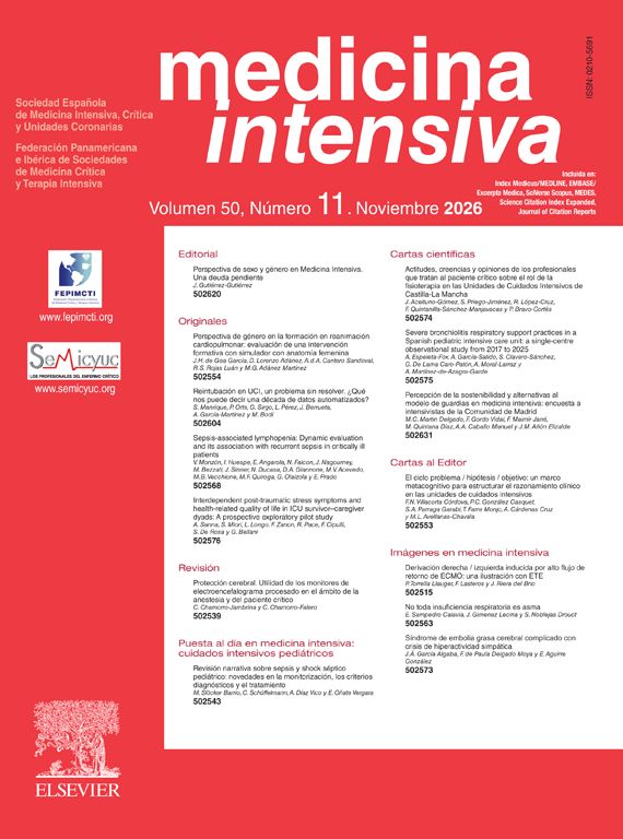
Protección cerebral. Utilidad de los monitores de electroencefalograma procesado en el ámbito de la anestesia y del paciente crítico

Neuroprotection. Usefulness of processed electroencephalogram monitors in anesthesia and critical care
Visitas
1340
Carlos Chamorro-Jambrinaa,
Autor para correspondencia
carlos.chamorro@salud.madrid.org

Autor para correspondencia.
, Carlos Chamorro-Falerob
a Servicio de Medicina Intensiva, HU Puerta de Hierro-Majadahonda, Majadahonda, Madrid, España
b Servicio de Anestesia, Hospital Universitario Virgen del Rocío, Sevilla, España
Este artículo ha recibido
Información del artículo
Resumen
Texto completo
Bibliografía
Descargar PDF
Estadísticas
Suplemento especial
Este artículo forma parte de:
El paciente neurocrítico

Editado por: el Dr. Juan Antonio Llompart Pou
Médico especialista en Medicina Intensiva, Doctor en Ciencias de la Salud por la Universitat Illes Balears. Servicio de Medicina Intensiva, UCI Trauma y Neurocríticos, Hospital Universitari Son Espases. Palma, España.

Última actualización: Octubre 2026

Más datos
Resumen

La monitorización electroencefalográfica procesada se ha consolidado como una herramienta clave para integrar la función cerebral en el manejo del paciente quirúrgico y crítico. La información combinada del EEG crudo, los índices procesados y la matriz de densidad espectral permite guiar la profundidad anestésica, optimizar la sedación y detectar precozmente estados de depresión cortical excesiva, contribuyendo a una estrategia de protección cerebral. Su uso facilita la identificación de cerebros vulnerables, la prevención del delirium y la individualización del tratamiento. Asimismo, permite reconocer actividad epiléptica subclínica, alteraciones de perfusión cerebral y cambios metabólicos, favoreciendo intervenciones tempranas. La interpretación debe realizarse de forma dinámica e integrada con el contexto clínico, ya que múltiples factores pueden modificar los patrones electroencefalográficos. Aunque presenta limitaciones técnicas y no sustituye al EEG convencional, su incorporación a la práctica mejora la seguridad, la toma de decisiones y promueve una monitorización cerebral personalizada en anestesia y cuidados intensivos.

Palabras clave:
Electroencefalograma procesado
Anestesia
Sedación
Paciente crítico
Conexión tálamo / cortical
Delirium
Deterioro cognitivo
Índice biespectral
Supresión electroencefalográfica
Estado epiléptico
Abstract

Processed electroencephalographic monitoring has become established as a key tool for integrating brain function into the management of surgical and critically ill patients. The combined information from the raw EEG, processed indices, and the spectral density matrix helps guide anesthetic depth, optimize sedation, and enable early detection of states of excessive cortical depression, contributing to a neuroprotective strategy. Its use facilitates the identification of vulnerable brains, the prevention of delirium, and the individualization of treatment. It also allows recognition of subclinical epileptic activity, alterations in cerebral perfusion, and metabolic changes, favoring early interventions. Interpretation should be performed dynamically and integrated with the clinical context, as multiple factors can modify electroencephalographic patterns. Although it has technical limitations and does not replace conventional EEG, its incorporation into practice improves safety and decision-making and promotes personalized brain monitoring in anesthesia and intensive care.

Keywords:
Processed electroencephalogram
Anesthesia
Sedation
Critically ill patient
Thalamocortical connectivity
Delirium
Cognitive impairment
Bispectral index
Electroencephalographic suppression
Status epilepticus

Artículo

Opciones para acceder a los textos completos de la publicación Medicina Intensiva
Socio
Si es usted socio de la Sociedad Española de Medicina Intensiva, Crítica y Unidades Coronarias:
Diríjase al área de socios de la web de la SEMICYUC (www.semicyuc.org ) y pulse el enlace a la revista.

Suscriptor
Suscriptor de la revista

Si ya tiene sus datos de acceso, clique aquí.

Si olvidó su clave de acceso puede recuperarla clicando aquí y seleccionando la opción "He olvidado mi contraseña".
Suscribirse
Suscribirse a:
Medicina Intensiva
Comprar
Comprar acceso al artículo

Comprando el artículo el PDF del mismo podrá ser descargado

Comprar ahora
Contactar
Teléfono para suscripciones e incidencias
De lunes a viernes de 9h a 18h (GMT+1) excepto los meses de julio y agosto que será de 9 a 15h
Llamadas desde España
932 415 960
Llamadas desde fuera de España
+34 932 415 960
Email
Bibliografía
[1]
S. Deiner, X. Liu, H.M. Lin, et al.
Does postoperative cognitive decline result in new disability after surgery?.
Ann Surg, 274 (2021), pp. e1108-e1114
[2]
L. Li, D. Dohan, A.K. Smith, E.L. Whitlock.
’It was a great brain, and I miss it’: Lay perspectives on postoperative cognitive dysfunction.
Br J Anaesth, 130 (2023), pp. 567-572
[3]
M.S. Herridge, É. Azoulay.
Outcomes after critical illness.
N Engl J Med, 388 (2023), pp. 913-924
[4]
C. Chamorro-Falero, J. García-García, J.J. Morales-Domene.
Delirium en el paciente posquirúrgico.
Rev Esp Anestesiol Reanim, 73 (2026),
[5]
N.E. Brummel, J.C. Jackson, P.P. Pandharipande, et al.
Delirium in the ICU and subsequent long-term disability among survivors of mechanical ventilation.
Crit Care Med, 42 (2014), pp. 369-377
[6]
S. Vacas, D.J. Cole, M. Cannesson.
Cognitive decline associated with anesthesia and surgery in older patients.
JAMA, 326 (2021), pp. 863-864
[7]
C. Giménez-Esparza, Relucio MÁ, K.L. Nanwani-Nanwani, J.M. Añón.
Impact of patient safety on outcomes. From prevention to the treatment of post-intensive care syndrome.
Med Intensiva (Engl Ed), 49 (2025), pp. 224-236
[8]
S. Alcántara Carmona, C. Chamorro-Jambrina.
Current role of midazolam in sedation of the ventilated critically ill patient: In favour.
Med Intensiva (Engl Ed), 48 (2024), pp. 174-176
[9]
G. Chanques, J.P. Kress.
Delirium related to sedation: Prevention strategies and patient perspectives.
Intensive Care Med, 51 (2025), pp. 1894-1897
[10]
A. Zare-Kaseb, N. Sanaie, S. Sarmadi.
Prevalence and incidence of post-intensive care syndrome among intensive care unit survivors: A systematic review and meta-analysis.
[11]
J. Caballero, M. García-Sánchez, E. Palencia-Herrejón, et al.
Oversedation Zero as a tool for comfort, safety and intensive care unit management.
Med Intensiva (Engl Ed), 44 (2020), pp. 239-247
[12]
T.G. Fong, S.K. Inouye.
The inter-relationship between delirium and dementia: The importance of delirium prevention.
Nat Rev Neurol, 18 (2022), pp. 579-596
[13]
V. Bonhomme, C. Putensen, B.W. Böttiger, et al.
Brain health: A concern for anaesthesiologists and intensivists.
Eur J Anaesthesiol Intensive Care, 3 (2024), pp. e0063
[14]
D. Friedman, J. Claassen, L.J. Hirsch.
Continuous electroencephalogram monitoring in the intensive care unit.
Anesth Analg, 109 (2009), pp. 506-523
[15]
N. Manohara, A. Ferrari, A. Greenblatt, et al.
Electroencephalogram monitoring during anesthesia and critical care: A guide for the clinician.
J Clin Monit Comput, 39 (2025), pp. 315-348
[16]
A. Alkhachroum, B. Appavu, S. Egawa, et al.
Electroencephalogram in the intensive care unit: A focused look at acute brain injury.
Intensive Care Med, 48 (2022), pp. 1443-1462
[17]
S. Rajagopalan, A. Sarwal.
Neuromonitoring in critically ill patients.
Crit Care Med, 51 (2023), pp. 525-542
[18]
P.L. Purdon, A. Sampson, K.J. Pavone, E.N. Brown.
Clinical electroencephalography for anesthesiologists: Part I: background and basic signatures.
Anesthesiology, 123 (2015), pp. 937-960
[19]
S. Romagnoli, F. Franchi, Z. Ricci.
Processed EEG monitoring for anesthesia and intensive care practice.
Minerva Anestesiol, 85 (2019), pp. 1219-1230
[20]
F. Iturri Clavero.
Monitorización perioperatoria de la función cerebral, mucho más que «hipnometría».
Rev Esp Anestesiol Reanim, 70 (2023), pp. S34-S45
[21]
C. Dubost, P. Humbert, A. Benizri, J.P. Tourtier, N. Vayatis, P.P. Vidal.
Selection of the best electroencephalogram channel to predict the depth of anesthesia.
Front Comput Neurosci, 13 (2019), pp. 65
[22]
C.S. Guay, U. Agrawal, B. Tseng, S. Gallo, D.R. Schreier, E.N. Brown.
Clinical electroencephalography for anesthesiologists and intensivists: Part 2-physiologic signatures and active management.
Anesthesiology, 143 (2025), pp. 1595-1618
[23]
S. Vijayan, S. Ching, P.L. Purdon, E.N. Brown, N.J. Kopell.
Thalamocortical mechanisms for the anteriorization of α rhythms during propofol-induced unconsciousness.
J Neurosci, 33 (2013), pp. 11070-11075
[24]
E.N. Brown, R. Lydic, N.D. Schiff.
General anesthesia, sleep, and coma.
N Engl J Med, 363 (2010), pp. 2638-2650
[25]
S. Alcántara Carmona, Romera Ortega MÁ, C. Chamorro-Jambrina.
Bispectral index monitoring vs clinical assessment for deep sedation in the ICU: It's not all about the number.
Chest, 167 (2025), pp. e101-e102
[26]
P.S. García, P.A. Goldstein.
The myth of depth of anesthesia monitors: when a “normal” bispectral index does not mean what you think it means.
Anesth Analg, 142 (2026), pp. 1050-1054
[27]
I.J. Rampil.
A primer for EEG signal processing in anesthesia.
Anesthesiology, 89 (1998), pp. 980-1002
[28]
D. Hight, M. Kreuzer, G. Ugen, et al.
Five commercial “depth of anaesthesia” monitors provide discordant clinical recommendations in response to identical emergence-like EEG signals.
Br J Anaesth, 130 (2023), pp. 536-545
[29]
J. Montupil, A. Defresne, V. Bonhomme.
The raw and processed electroencephalogram as a monitoring and diagnostic tool.
J Cardiothorac Vasc Anesth, 33 (2019), pp. S3-S10
[30]
A. Fleischmann, M.T. Georgii, J. Schuessler, G. Schneider, S. Pilge, M. Kreuzer.
Always assess the raw electroencephalogram: Why automated burst suppression detection may not detect all episodes.
Anesth Analg, 136 (2023), pp. 346-354
[31]
P.L. Purdon, E.T. Pierce, E.A. Mukamel, et al.
Electroencephalogram signatures of loss and recovery of consciousness from propofol.
Proc Natl Acad Sci U S A, 110 (2013), pp. E1142-E1151
[32]
M. Malekmohammadi, C.M. Price, A.E. Hudson, J.A.T. DiCesare, N. Pouratian.
Propofol-induced loss of consciousness is associated with a decrease in thalamocortical connectivity in humans.
Brain, 142 (2019), pp. 2288-2302
[33]
J.A. Stewart, M.O.K. Särkelä, J. Wennervirta, A.P. Vakkuri.
Novel insights on association and reactivity of bispectral Index, frontal electromyogram, and autonomic responses in nociception-sedation monitoring of critical care patients.
BMC Anesthesiol, 22 (2022), pp. 353
[34]
P.S. García, M. Kreuzer, D. Hight, J.W. Sleigh.
Effects of noxious stimulation on the electroencephalogram during general anaesthesia: A narrative review and approach to analgesic titration.
Br J Anaesth, 126 (2021), pp. 445-457
[35]
K.K. MacKenzie, A.M. Britt-Spells, L.P. Sands, J.M. Leung.
Processed electroencephalogram monitoring and postoperative delirium: A systematic review and meta-analysis.
Anesthesiology, 129 (2018), pp. 417-427
[36]
A. Xu, B. Arnaout, D.A. Scott, S. McGuigan.
Intraoperative electroencephalogram-derived measures and their association with postoperative delirium: A systematic review and meta-analysis.
Br J Anaesth, 135 (2025), pp. 1684-1703
[37]
M. Sumner, C. Deng, L. Evered, et al.
Processed electroencephalography-guided general anaesthesia to reduce postoperative delirium: A systematic review and meta-analysis.
Br J Anaesth, 130 (2023), pp. e243-e253
[38]
S.K. Park, D.W. Han, C.H. Chang, H. Jung, H. Kang, Y. Song.
Association between intraoperative electroencephalogram burst suppression and postoperative delirium: A systematic review and meta-analysis.
Anesthesiology, 142 (2025), pp. 107-120
[39]
J.M. Andresen, T.D. Girard, P.P. Pandharipande, M.A. Davidson, E.W. Ely, P.L. Watson.
Burst suppression on processed electroencephalography as a predictor of postcoma delirium in mechanically ventilated ICU patients.
Crit Care Med, 42 (2014), pp. 2244-2251
[40]
P.L. Watson, A.K. Shintani, R. Tyson, P.P. Pandharipande, B.T. Pun, E.W. Ely.
Presence of electroencephalogram burst suppression in sedated, critically ill patients is associated with increased mortality.
Crit Care Med, 36 (2008), pp. 3171-3177
[41]
J. Hogan, H. Sun, H. Aboul Nour, et al.
Burst suppression: Causes and effects on mortality in critical illness.
Neurocrit Care, 33 (2020), pp. 565-574
[42]
R. Gutierrez, J.I. Egaña, I. Saez, et al.
Intraoperative low alpha power in the electroencephalogram is associated with postoperative subsyndromal delirium.
Front Syst Neurosci, 13 (2019), pp. 56
[43]
D. Hight, A. Ehrhardt, F. Lersch, M.M. Luedi, F. Stüber, H.A. Kaiser.
Lower alpha frequency of intraoperative frontal EEG is associated with postoperative delirium: A secondary propensity-matched analysis.
[44]
M. Carretero-Rodrigo, A. Amaro-Harpigny, E. Bueso-Inchausti, M.A. Romera-Ortega, Chamorro Jambrina.
Estudio ADELPHA. Asociación entre delirium y presencia de banda alfa en el EEG procesado durante la sedación profunda.
Med Intensiva, 49 (2025), pp. 186
[45]
S. Hesse, M. Kreuzer, D. Hight, et al.
Association of electroencephalogram trajectories during emergence from anaesthesia with delirium in the postanaesthesia care unit: An early sign of postoperative complications.
Br J Anaesth, 122 (2019), pp. 622-634
[46]
S. Dragovic, G. Schneider, P.S. García, et al.
Predictors of low risk for delirium during anesthesia emergence.
Anesthesiology, 139 (2023), pp. 757-768
[47]
C. Chamorro-Jambrina, C. Chamorro-Falero.
Don’t turn-off the brain [Article in English, Spanish].
Med Intensiva (Engl Ed), 43 (2019), pp. 1-2
[48]
M. Lamperti, F.A. Lobo.
The silent line: confronting iatrogenic cerebral suppression in the perioperative and critical care settings.
Intensive Care Med, (2025 Dec 22),
[49]
L.J. Hirsch, M.T. Gopaul.
EEG in the critical care setting.
[50]
C. Chamorro, M.A. Romera, B. Balandín, M. Valdivia.
Nonconvulsive status and bispectral index.
Crit Care Med, 36 (2008), pp. 2218-2219
[51]
L.J. Hirsch, M.W.K. Fong, M. Leitinger, et al.
American clinical neurophysiology society's standardized critical care EEG terminology: 2021 Version.
J Clin Neurophysiol, 38 (2021), pp. 1-29
[52]
S.F. Zafar, E. Amorim, C.A. Williamsom, et al.
A standardized nomenclature for spectrogram EEG patterns: Inter-rater agreement and correspondence with common intensive care unit EEG patterns.
Clin Neurophysiol, 131 (2020), pp. 2298-2306
[53]
M.A. Hernández-Hernández, J.L. Fernández-Torre.
Color density spectral array of bilateral bispectral index system: electroencephalographic correlate in comatose patients with nonconvulsive status epilepticus.
[54]
M.S. Cherchi, M.A. Hernández-Hernández, J. Hernández-Cabello, et al.
Routine EEG with quantitative analysis to detect delayed cerebral ischemia in aneurysmal subarachnoid hemorrhage.
[55]
C. Duclos, C. Maschke, Y. Mahdid, et al.
Brain responses to propofol in advance of recovery from coma and disorders of consciousness: A preliminary study.
Am J Respir Crit Care Med, 205 (2022), pp. 171-182
[56]
M.S. Avidan, G.A. Mashour.
Repurposing propofol as a prognostic probe for return of consciousness.
Am J Respir Crit Care Med, 205 (2022), pp. 140-142
[57]
E. Adam, O. Kwon, K.A. Montejo, E.N. Brown.
Modulatory dynamics mark the transition between anesthetic states of unconsciousness.
Proc Natl Acad Sci U S A, 120 (2023),
[58]
A. Rodrigues, C. Subirà, A. Bizios, et al.
Sedation-related electroencephalographic patterns in acute hypoxemic respiratory failure.
Anesthesiology, 143 (2025), pp. 1266-1278
[59]
F.A. Lobo, C. Robba, M. Lamperti, S. Romagnoli, F.A. Rasulo.
Processed EEG monitoring in critical care: A black swan or a shining star?.
J Clin Monit Comput, 37 (2023), pp. 3-5
[60]
F.A. Rasulo, P. Hopkins, F.A. Lobo, et al.
Processed electroencephalogram-based monitoring to guide sedation in critically ill adult patients: Recommendations from an international expert panel-based consensus.
Neurocrit Care, 38 (2023), pp. 296-311
Idiomas
Medicina Intensiva
Opciones de artículo
Herramientas