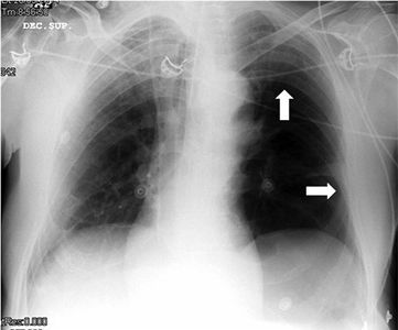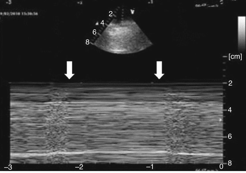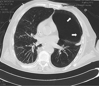


Varón de 71 años con criterios de EPOC grave (FEV1 38%), que ingresa en la UCI por insuficiencia respiratoria hipoxémica grave secundaria a infección respiratoria iniciándose antibioterapia empírica, tratamiento broncodilatador y ventilación mecánica invasiva. En la radiografía de tórax tras intubación (fig. 1) se objetiva imagen compatible con neumotórax izquierdo y se coloca tubo de tórax lateral sin fuga evidente, por lo que realizamos ecografía con signos de persistencia del neumotórax: líneas A, punto pulmonar (fig. 2) y ausencia de deslizamiento pleural. Ante la duda clínica se solicita TC torácica (fig. 3) en la que se identifica una imagen radiolucente redondeada de 10×8,5cm con pared fina sugestiva de ampolla anterior, sin línea clara de neumotórax. En pacientes con enfisema bulloso la ecografía pulmonar puede perder especificidad en el diagnóstico de neumotórax.