Article
Go to the members area of the website of the SEMICYUC (www.semicyuc.org )and click the link to the magazine.
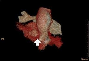
array:23 [ "pii" => "S2173572724000511" "issn" => "21735727" "doi" => "10.1016/j.medine.2024.02.014" "estado" => "S200" "fechaPublicacion" => "2024-03-21" "aid" => "1990" "copyright" => "Elsevier España, S.L.U. and SEMICYUC" "copyrightAnyo" => "2024" "documento" => "article" "crossmark" => 0 "subdocumento" => "sco" "abierto" => array:3 [ "ES" => false "ES2" => false "LATM" => false ] "gratuito" => false "lecturas" => array:1 [ "total" => 0 ] "Traduccion" => array:1 [ "es" => array:18 [ "pii" => "S0210569124000676" "issn" => "02105691" "doi" => "10.1016/j.medin.2024.02.004" "estado" => "S200" "fechaPublicacion" => "2024-03-16" "aid" => "1990" "copyright" => "Elsevier España, S.L.U. y SEMICYUC" "documento" => "article" "crossmark" => 0 "subdocumento" => "sco" "abierto" => array:3 [ "ES" => false "ES2" => false "LATM" => false ] "gratuito" => false "lecturas" => array:1 [ "total" => 0 ] "es" => array:10 [ "idiomaDefecto" => true "cabecera" => "<span class="elsevierStyleTextfn">Imágenes en Medicina Intensiva</span>" "titulo" => "Perforación de base de cráneo por cocaína" "tienePdf" => "es" "tieneTextoCompleto" => "es" "titulosAlternativos" => array:1 [ "en" => array:1 [ "titulo" => "Base skull perforation due to cocaine" ] ] "contieneTextoCompleto" => array:1 [ "es" => true ] "contienePdf" => array:1 [ "es" => true ] "resumenGrafico" => array:2 [ "original" => 0 "multimedia" => array:6 [ "identificador" => "fig0005" "etiqueta" => "Figura 1" "tipo" => "MULTIMEDIAFIGURA" "mostrarFloat" => true "mostrarDisplay" => false "figura" => array:1 [ 0 => array:4 [ "imagen" => "gr1.jpeg" "Alto" => 940 "Ancho" => 1255 "Tamanyo" => 169799 ] ] ] ] "autores" => array:1 [ 0 => array:2 [ "autoresLista" => "Juan Maria Romeu Prieto, María Teresa Correas López, Jhoann Miguel Guerra Cuervo" "autores" => array:3 [ 0 => array:2 [ "nombre" => "Juan Maria" "apellidos" => "Romeu Prieto" ] 1 => array:2 [ "nombre" => "María Teresa" "apellidos" => "Correas López" ] 2 => array:2 [ "nombre" => "Jhoann Miguel" "apellidos" => "Guerra Cuervo" ] ] ] ] ] "idiomaDefecto" => "es" "Traduccion" => array:1 [ "en" => array:9 [ "pii" => "S2173572724000511" "doi" => "10.1016/j.medine.2024.02.014" "estado" => "S200" "subdocumento" => "" "abierto" => array:3 [ "ES" => false "ES2" => false "LATM" => false ] "gratuito" => false "lecturas" => array:1 [ "total" => 0 ] "idiomaDefecto" => "en" "EPUB" => "https://multimedia.elsevier.es/PublicationsMultimediaV1/item/epub/S2173572724000511?idApp=WMIE" ] ] "EPUB" => "https://multimedia.elsevier.es/PublicationsMultimediaV1/item/epub/S0210569124000676?idApp=WMIE" "url" => "/02105691/unassign/S0210569124000676/v1_202403161144/es/main.assets" ] ] "itemSiguiente" => array:17 [ "pii" => "S2173572724000481" "issn" => "21735727" "doi" => "10.1016/j.medine.2024.02.012" "estado" => "S200" "fechaPublicacion" => "2024-03-25" "aid" => "1984" "documento" => "article" "crossmark" => 0 "subdocumento" => "sco" "abierto" => array:3 [ "ES" => false "ES2" => false "LATM" => false ] "gratuito" => false "lecturas" => array:1 [ "total" => 0 ] "en" => array:10 [ "idiomaDefecto" => true "cabecera" => "<span class="elsevierStyleTextfn">Images in Intensive Medicine</span>" "titulo" => "Esophageal perforation secondary to achalasia" "tienePdf" => "en" "tieneTextoCompleto" => "en" "titulosAlternativos" => array:1 [ "es" => array:1 [ "titulo" => "Perforación esofágica secundaria a una acalasia" ] ] "contieneTextoCompleto" => array:1 [ "en" => true ] "contienePdf" => array:1 [ "en" => true ] "resumenGrafico" => array:2 [ "original" => 0 "multimedia" => array:7 [ "identificador" => "fig0010" "etiqueta" => "Figure 2" "tipo" => "MULTIMEDIAFIGURA" "mostrarFloat" => true "mostrarDisplay" => false "figura" => array:1 [ 0 => array:4 [ "imagen" => "gr2.jpeg" "Alto" => 614 "Ancho" => 680 "Tamanyo" => 52864 ] ] "detalles" => array:1 [ 0 => array:3 [ "identificador" => "at0010" "detalle" => "Figure " "rol" => "short" ] ] ] ] "autores" => array:1 [ 0 => array:2 [ "autoresLista" => "Rosa María Vela Colmenero, Crispín Colmenero Aguilar, Ana María Vela Colmenero" "autores" => array:3 [ 0 => array:2 [ "nombre" => "Rosa María" "apellidos" => "Vela Colmenero" ] 1 => array:2 [ "nombre" => "Crispín" "apellidos" => "Colmenero Aguilar" ] 2 => array:2 [ "nombre" => "Ana María" "apellidos" => "Vela Colmenero" ] ] ] ] ] "idiomaDefecto" => "en" "Traduccion" => array:1 [ "es" => array:9 [ "pii" => "S0210569124000597" "doi" => "10.1016/j.medin.2024.01.012" "estado" => "S200" "subdocumento" => "" "abierto" => array:3 [ "ES" => false "ES2" => false "LATM" => false ] "gratuito" => false "lecturas" => array:1 [ "total" => 0 ] "idiomaDefecto" => "es" "EPUB" => "https://multimedia.elsevier.es/PublicationsMultimediaV1/item/epub/S0210569124000597?idApp=WMIE" ] ] "EPUB" => "https://multimedia.elsevier.es/PublicationsMultimediaV1/item/epub/S2173572724000481?idApp=WMIE" "url" => "/21735727/unassign/S2173572724000481/v1_202403250352/en/main.assets" ] "itemAnterior" => array:17 [ "pii" => "S2173572724000274" "issn" => "21735727" "doi" => "10.1016/j.medine.2024.02.003" "estado" => "S200" "fechaPublicacion" => "2024-02-25" "aid" => "1976" "copyright" => "Elsevier España, S.L.U. and SEMICYUC" "documento" => "article" "crossmark" => 0 "subdocumento" => "fla" "abierto" => array:3 [ "ES" => false "ES2" => false "LATM" => false ] "gratuito" => false "lecturas" => array:1 [ "total" => 0 ] "en" => array:12 [ "idiomaDefecto" => true "cabecera" => "<span class="elsevierStyleTextfn">Original article</span>" "titulo" => "Fractional excretion of sodium and potassium and urinary strong ion difference in the evaluation of persistent AKI in sepsis" "tienePdf" => "en" "tieneTextoCompleto" => "en" "tieneResumen" => array:2 [ 0 => "en" 1 => "es" ] "titulosAlternativos" => array:1 [ "es" => array:1 [ "titulo" => "Excreción fraccional de sodio y potasio, y brecha aniónica urinaria en la evaluación de la IRA persistente en sepsis" ] ] "contieneResumen" => array:2 [ "en" => true "es" => true ] "contieneTextoCompleto" => array:1 [ "en" => true ] "contienePdf" => array:1 [ "en" => true ] "resumenGrafico" => array:2 [ "original" => 0 "multimedia" => array:8 [ "identificador" => "fig0010" "etiqueta" => "Figure 2" "tipo" => "MULTIMEDIAFIGURA" "mostrarFloat" => true "mostrarDisplay" => false "figura" => array:1 [ 0 => array:4 [ "imagen" => "gr2.jpeg" "Alto" => 1448 "Ancho" => 1675 "Tamanyo" => 110835 ] ] "detalles" => array:1 [ 0 => array:3 [ "identificador" => "at0135" "detalle" => "Figure " "rol" => "short" ] ] "descripcion" => array:1 [ "en" => "<p id="spar0015" class="elsevierStyleSimplePara elsevierViewall">ROC curve showing the area under de curve of FENa, FEK and uSID.</p> <p id="spar0020" class="elsevierStyleSimplePara elsevierViewall">FENa: fractional excretion of sodium; FEK: fractional excretion of potassium; uSID: urinary strong ion difference.</p>" ] ] ] "autores" => array:1 [ 0 => array:2 [ "autoresLista" => "Nicolás Contrera Rolón, Joaquín Cantos, Iván Huespe, Eduardo Prado, Griselda I. Bratti, Carlos Schreck, Sergio Giannasi, Guillermo Rosa Diez, Carlos F. Varela" "autores" => array:9 [ 0 => array:2 [ "nombre" => "Nicolás" "apellidos" => "Contrera Rolón" ] 1 => array:2 [ "nombre" => "Joaquín" "apellidos" => "Cantos" ] 2 => array:2 [ "nombre" => "Iván" "apellidos" => "Huespe" ] 3 => array:2 [ "nombre" => "Eduardo" "apellidos" => "Prado" ] 4 => array:2 [ "nombre" => "Griselda I." "apellidos" => "Bratti" ] 5 => array:2 [ "nombre" => "Carlos" "apellidos" => "Schreck" ] 6 => array:2 [ "nombre" => "Sergio" "apellidos" => "Giannasi" ] 7 => array:2 [ "nombre" => "Guillermo" "apellidos" => "Rosa Diez" ] 8 => array:2 [ "nombre" => "Carlos F." "apellidos" => "Varela" ] ] ] ] ] "idiomaDefecto" => "en" "EPUB" => "https://multimedia.elsevier.es/PublicationsMultimediaV1/item/epub/S2173572724000274?idApp=WMIE" "url" => "/21735727/unassign/S2173572724000274/v1_202402251056/en/main.assets" ] "en" => array:11 [ "idiomaDefecto" => true "cabecera" => "<span class="elsevierStyleTextfn">Images in Intensive Medicine</span>" "titulo" => "Perforation of the skull base due to cocaine" "tieneTextoCompleto" => true "autores" => array:1 [ 0 => array:4 [ "autoresLista" => "Juan María Romeu Prieto, María Teresa Correas López, Jhoann Miguel Guerra Cuervo" "autores" => array:3 [ 0 => array:2 [ "nombre" => "Juan María" "apellidos" => "Romeu Prieto" ] 1 => array:2 [ "nombre" => "María Teresa" "apellidos" => "Correas López" ] 2 => array:4 [ "nombre" => "Jhoann Miguel" "apellidos" => "Guerra Cuervo" "email" => array:1 [ 0 => "jmromeu@sescam.jccm.es" ] "referencia" => array:1 [ 0 => array:2 [ "etiqueta" => "*" "identificador" => "cor0005" ] ] ] ] "afiliaciones" => array:1 [ 0 => array:3 [ "entidad" => "Complejo Hospitalario Universitario de Toledo, Toledo, Spain" "etiqueta" => "a" "identificador" => "aff0005" ] ] "correspondencia" => array:1 [ 0 => array:3 [ "identificador" => "cor0005" "etiqueta" => "⁎" "correspondencia" => "Corresponding author." ] ] ] ] "titulosAlternativos" => array:1 [ "es" => array:1 [ "titulo" => "Perforación de base de cráneo por cocaína" ] ] "resumenGrafico" => array:2 [ "original" => 0 "multimedia" => array:7 [ "identificador" => "fig0005" "etiqueta" => "Figure 1" "tipo" => "MULTIMEDIAFIGURA" "mostrarFloat" => true "mostrarDisplay" => false "figura" => array:1 [ 0 => array:4 [ "imagen" => "gr1.jpeg" "Alto" => 940 "Ancho" => 1255 "Tamanyo" => 169814 ] ] "detalles" => array:1 [ 0 => array:3 [ "identificador" => "at0005" "detalle" => "Figure " "rol" => "short" ] ] ] ] "textoCompleto" => "<span class="elsevierStyleSections"><p id="par0005" class="elsevierStylePara elsevierViewall">A 50-year-old woman reported to the emergency room due to walking instability, right-side body paresis and diplopia with complete involvement of the right third cranial nerve. As to her medical history, the main observation was regular cocaine use for the last 20 years.</p><p id="par0010" class="elsevierStylePara elsevierViewall">The brain computed tomography scan revealed clivus perforation (white arrow). The magnetic resonance imaging scan in turn also evidenced involvement of the odontoid process (yellow circle and arrow), consistent with regular cocaine use, with an associated abscess that compressed the pons and bulb of the mesencephalon.</p><p id="par0015" class="elsevierStylePara elsevierViewall">Cocaine is known to cause destruction due to vasoconstriction and necrosis of the nasal septum and hard palate, but its effects on the skull base and even the cervical vertebras have been less widely described (<a class="elsevierStyleCrossRef" href="#fig0005">Fig. 1</a>).</p><elsevierMultimedia ident="fig0005"></elsevierMultimedia></span>" "pdfFichero" => "main.pdf" "tienePdf" => true "multimedia" => array:1 [ 0 => array:7 [ "identificador" => "fig0005" "etiqueta" => "Figure 1" "tipo" => "MULTIMEDIAFIGURA" "mostrarFloat" => true "mostrarDisplay" => false "figura" => array:1 [ 0 => array:4 [ "imagen" => "gr1.jpeg" "Alto" => 940 "Ancho" => 1255 "Tamanyo" => 169814 ] ] "detalles" => array:1 [ 0 => array:3 [ "identificador" => "at0005" "detalle" => "Figure " "rol" => "short" ] ] ] ] ] "idiomaDefecto" => "en" "url" => "/21735727/unassign/S2173572724000511/v1_202403210441/en/main.assets" "Apartado" => null "PDF" => "https://static.elsevier.es/multimedia/21735727/unassign/S2173572724000511/v1_202403210441/en/main.pdf?idApp=WMIE&text.app=https://www.medintensiva.org/" "EPUB" => "https://multimedia.elsevier.es/PublicationsMultimediaV1/item/epub/S2173572724000511?idApp=WMIE" ]