The pharmacological group of immune checkpoint-inhibitors (ICI) has revolutionized the field of oncology in the last ten years. The improvements in the survival of certain cancers thanks to these treatments comes at the cost of an increased morbidity and mortality due to certain immune related adverse events (irAE). This review will concentrate on the irAE that more frequently require intensive care unit (ICU) admission. The infectious burden of patients treated with ICI is also explored, shining light not only on the infections caused by the immunosuppression needed to manage the different irAE, but also on the specific infections arising from a unique immune dysregulation only seen in ICI treated patients.
El grupo farmacológico de los checkpoint-inhibitors (ICI) ha revolucionado la oncología en los últimos diez años. Las mejorías en el pronóstico oncológico se han acompañado de una serie de eventos adversos inmunomediados (irAE) que otorgan importante morbi-mortalidad. Esta revisión está centrada en los irAE que con mayor frecuencia pueden requerir un ingreso en una unidad de cuidados intensivos (UCI). Además, aporta un énfasis en la importancia de la patología infecciosa que pueden sufrir estos enfermos, ya sea por la inmunosupresión requerida para tratar los irAE, como por la disregulación inmune propia de los ICI.
Immune checkpoint inhibitors (ICI) are a group of humanized monoclonal antibodies that have revolutionized the field of oncology since Ipilimumab (the first member of this group), was approved for the treatment of melanoma back in 2011.1 ICI have changed the overall survival of several advanced malignancies to the extent of sustained remissions, like in the case of malignant metastatic melanoma and Non-Small Cell Lung Cancer (NSCLC). By 2022, the FDA has approved a total of 9 different ICI used for the treatment of more than 15 different malignancies.
This benefit of survival comes at the cost of specific immune related adverse events (irAE). These side effects can affect the quality of life of patients and cause significant morbidity and mortality. Close to 50% of intensive care unit (ICU) admissions in cancer patients treated with ICI are motivated by these irAE.2
The main challenge when diagnosing irAE is ruling out an infectious complication. Infections represent the second most frequent cause for ICU admissions in ICI treated patients2 and the use of immunosuppression to manage irAE makes the risk even higher.
This review will focus on the diagnosis and management of the main immune mediated and infectious adverse events in patients treated with ICI.
Materials and methodsThis text is based on a PubMed review of the recent publications (from 2015 to 2024) on the topic of ICI based adverse events. We emphasized on the side effects that most frequently resulted in an ICU admission. The review also includes recommendations based on American and European guidelines of clinical oncology.
Mechanism of action and specific considerationsICI act by interfering with inhibitory signals between lymphocytes. These inhibitory signals represent the physiologic mechanisms by which the immune system modulates inflammatory responses and generates tolerance. The cancer microenvironment selects specific cellular clones that use these inhibitory signals in their advantage, making them resistant to the immune surveillance done by cytotoxic T lymphocytes. The proteins responsible for these inhibitory signals are called checkpoints. Some of them are: cytotoxic T cell antigen 4 (CTLA4), programmed death receptor (PD) and programmed death ligand (PD-L).
CTLA-4 is expressed on the Surface of activated CD4 lymphocytes and binds to CD80/CD86 on the surface of antigen presenting cells (APC) (Fig. 1). When bound to its ligands, CTLA-4 causes a downstream deactivation of CD4 lymphocytes. Ipilimumab is the only ICI targeting this checkpoint molecule. Its effects on derepressing naive CD4 lymphocytes generates a large clonal variety and expansion; this makes the immune effects of Ipilimumab long lasting, tumour recurrences less frequent and treatments shorter (usually 2–3 doses).
Sites of action of the different ICI used in clinical practice. + denotes cellular activation and – refers to cellular inhibition. TCR: T cell receptor. HLA: human leukocyte antigen. CD: cluster of differentiation. CTLA 4: cytotoxic T lymphocyte antigen 4. PD-1: programmed death receptor 1. PD-L1: programmed death receptor antigen.
Illustration made by the author, based on the text and inspired by the figures used int the article by MareI et al.1
PD-1 receptors are expressed on the surface of activated CD4 and CD8 T lymphocytes. Their ligands, PD-L1 and PD-L2 are expressed in a large variety of cells across the body (Fig. 1).3 When PD-L1 is expressed on the surface of a cell, its binding to PD-1 positive CD8 lymphocytes causes T cell deactivation and tolerance. Over expression ofPD-L by cancer cells generates a large amount of senescent CD8+ lymphocytes. Inhibition of PD1/PD1-L signalling cascade by specific ICI derepresses the acquired peripheral tissue and cancer tolerance. This second group of ICIs tend to have a shorter immunologic effect with a higher tumoral recurrence and longer treatment regimes, usually 2 years of monthly dosages.
More recently, combination regimens of anti-CTLA-4 and anti-PD1/PD-L1 have proven to be superior in terms of survival in several tumour types but at the cost of more severe irAE. In one observational study by Lin et al.,4 up to 63% of the irAE requiring ICU admission were related to combination regimens of ipilimumab¿;+¿;nivolumab.
When talking about irAE, most literature refers to organ disfunctions mediated by the immune system. The most frequent pathological finding associated with irAE is an extensive tissue infiltration by T lymphocytes (mainly CD8+) whereas humoral immunity and B lymphocytes seem to play a smaller role.5
Although in recent years there has been a growing concern about a different kind of irAE, the ones caused by enhanced immunity against infective pathogens, what Morelli et al.6 called immunotherapy infections due to dysregulated immunity (ITI-DI). These second kind of adverse events manifest as infections produced by cytomegalovirus (CMV), typical and atypical mycobacteria and invasive pulmonary aspergillosis (IPA), among others. They can appear even in the absence of immunosuppressive therapy (corticosteroids or Infliximab) and are thought to be caused by the maladaptive activation of the immune system against latent infections.
Immune related adverse eventsThe most relevant irAE that might require ICU admission will be reviewed in this section. Their appearance depends on:
Individual factors: having a good functional status measured by the Eastern Cooperative Oncology Group (ECOG) as 0–1 and a previous history of irAE were both associated with severe irAE requiring ICU admission.2
Specific agent factors: anti-CTLA4 generally causes colitis and anti-PD1/PD-L1 cause pneumonitis.
Tumor type: NSCLC has a higher risk for pneumonitis, melanoma carries a higher risk for peripheral neuropathy. The two most common tumour types associated with irAE related ICU admisions were precisely NSCLC and melanoma.2,4,7
The irAE can be classified in 5 grades (Table 1) depending on their severity.8 Grades 3 or more are considered severe and account for 1–10% of all the irAE of patients receiving ICI.9 Thankfully only a small proportion of patients with severe irAE require actual ICU admission, depending on the source this number ranges from 0,6 to 1,2%.4,7
Grading system of the severity of immune related adverse events. Based on the Common Terminology Criteria for Adverse Events (CTCAE).
irAE Grading | Grade 1 | Grade 2 | Grade 3 | Grade 4 | Grade 5 |
---|---|---|---|---|---|
Characteristics | Mild symptoms that do not interfere with daily activities | Moderate symptoms that do interfere with daily activities | Severe symptoms that require hospital admision. | Very severe symptoms that threaten patient's life | Symptoms that cause the death of the patient |
Management | No treatment needed | Treatment is needed | Treatment is needed | Treatment is needed | |
No interruption of ICI | Temporary interruption of ICI | Permanent interruption of ICI |
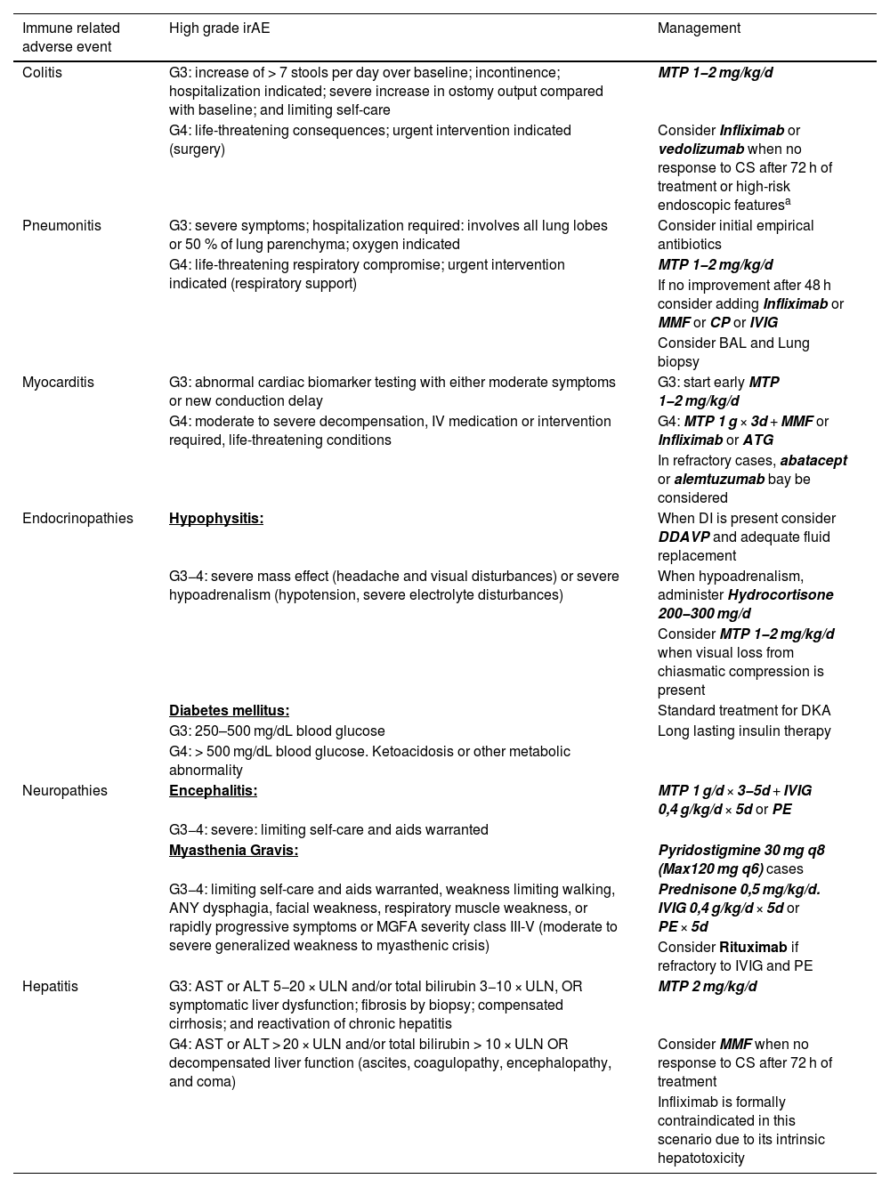
Patients with G4 irAE frequently require ICU admission and immunosuppressive treatment (Table 2). In the largest prospective study to date carried out by Toffart et al.2 the main cause of ICU admission in patients with suspected of irAE was respiratory failure (66,57%) followed by colitis (14,13%) and cardiovascular disease (13,11%). The overall ICU mortality in this study was 21%, with the highest ICU mortalities (28%) in patients admitted for respiratory failure. Another multicentre study by Joseph et al.10 showed that ICU mortality attributed to irAE was significantly lower than other admissions like sepsis (17,2% vs 23%; p¿;=¿;0,004). Other smaller studies7 support the overall good prognosis of irAE requiring ICU admission, especially when detection and treatment are done early.
Criteria that define the most severe forms of irAE and their management. Based on the American Society of Clinical Oncology (ASCO) guidelines.
Immune related adverse event | High grade irAE | Management |
---|---|---|
Colitis | G3: increase of > 7 stools per day over baseline; incontinence; hospitalization indicated; severe increase in ostomy output compared with baseline; and limiting self-care | MTP 1−2¿;mg/kg/d |
G4: life-threatening consequences; urgent intervention indicated (surgery) | Consider Infliximab or vedolizumab when no response to CS after 72¿;h of treatment or high-risk endoscopic featuresa | |
Pneumonitis | G3: severe symptoms; hospitalization required: involves all lung lobes or 50 % of lung parenchyma; oxygen indicated | Consider initial empirical antibiotics |
G4: life-threatening respiratory compromise; urgent intervention indicated (respiratory support) | MTP 1−2¿;mg/kg/d | |
If no improvement after 48¿;h consider adding Infliximab or MMF or CP or IVIG | ||
Consider BAL and Lung biopsy | ||
Myocarditis | G3: abnormal cardiac biomarker testing with either moderate symptoms or new conduction delay | G3: start early MTP 1−2¿;mg/kg/d |
G4: moderate to severe decompensation, IV medication or intervention required, life-threatening conditions | G4: MTP 1¿;g¿;׿;3d¿;+¿;MMF or Infliximab or ATG | |
In refractory cases, abatacept or alemtuzumab bay be considered | ||
Endocrinopathies | Hypophysitis: | When DI is present consider DDAVP and adequate fluid replacement |
G3−4: severe mass effect (headache and visual disturbances) or severe hypoadrenalism (hypotension, severe electrolyte disturbances) | When hypoadrenalism, administer Hydrocortisone 200−300¿;mg/d | |
Consider MTP 1−2¿;mg/kg/d when visual loss from chiasmatic compression is present | ||
Diabetes mellitus: | Standard treatment for DKA | |
G3: 250–500¿;mg/dL blood glucose | Long lasting insulin therapy | |
G4: > 500¿;mg/dL blood glucose. Ketoacidosis or other metabolic abnormality | ||
Neuropathies | Encephalitis: | MTP 1¿;g/d¿;׿;3−5d¿;+¿;IVIG 0,4¿;g/kg/d¿;׿;5d or PE |
G3−4: severe: limiting self-care and aids warranted | ||
Myasthenia Gravis: | Pyridostigmine 30¿;mg q8 (Max120¿;mg q6) cases | |
G3−4: limiting self-care and aids warranted, weakness limiting walking, ANY dysphagia, facial weakness, respiratory muscle weakness, or rapidly progressive symptoms or MGFA severity class III-V (moderate to severe generalized weakness to myasthenic crisis) | Prednisone 0,5¿;mg/kg/d. IVIG 0,4¿;g/kg/d¿;׿;5d or PE¿;׿;5d | |
Consider Rituximab if refractory to IVIG and PE | ||
Hepatitis | G3: AST or ALT 5−20¿;׿;ULN and/or total bilirubin 3−10¿;׿;ULN, OR symptomatic liver dysfunction; fibrosis by biopsy; compensated cirrhosis; and reactivation of chronic hepatitis | MTP 2¿;mg/kg/d |
G4: AST or ALT¿;>¿;20¿;׿;ULN and/or total bilirubin > 10¿;׿;ULN OR decompensated liver function (ascites, coagulopathy, encephalopathy, and coma) | Consider MMF when no response to CS after 72¿;h of treatment | |
Infliximab is formally contraindicated in this scenario due to its intrinsic hepatotoxicity |
MTP: methylprednisolone. CS: corticosteroids. MMF: mycophenolate-mofetil. CP: cyclophosphamide. IVIG: intravenous immunoglobulins. ATG: anti-thymocyte globulin. DI: diabetes insipidus. DDAVP: desmopressin. DKA: diabetic ketoacidosis. PE: plasma exchange.
It is important to note that irAE have a particular chronology of appearance (Fig. 2). This chronology is helpful when establishing a differential diagnosis of organ disfunctions in patients treated with ICI. But we must have in mind that in combinations of ICI like ipilimumab and nivolumab, irAE tend to appear earlier and have higher grades of severity. Lethal toxicities can occur as early as 15 days after combined ICI treatment is initiated (and arise past 40 days when a single ICI is used).9 On the other end of the spectrum, irAE have been documented to occur months and years after discontinuation of ICI treatment. This late onset irAE have been called DIRE (Delayed immune-related Events) and are of growing concern given their additional morbidity and mortality due to their frequent misdiagnosis and delayed treatment.11,12
Chronology of appearance of the different irAE. The severity is represented by shades of warmer colours. Note that different regimens have different severities and chronologies for the same type of irAE. C¿;+¿;P: combination of anti-CTLA4 and anti-PD1/PD-L1 agents.
Lastly, irAE tend to overlap, so patients can have a rash, colitis and pneumonitis at the same time.
- •
Enterocolitis:
Enterocolitis is the most frequent irAE. It usually appears four weeks after starting treatment, but it can show up earlier in combined regimens of ICI. Close to 30% of all patients treated with ICI will suffer from diarrhoea but only 5% of them would be diagnosed with colitis.9,13,14 One of the major risk factors of suffering this adverse event is receiving Ipilimumab. Up to 54% of patients treated with ipilimumab suffer from diarrhoea when compared to the 19% of those treated with anti-PD1/PD-L1, and about 20% of those treated with ipilimumab will be diagnosed with colitis. Individual risk factors include, medical history of inflammatory bowel disease (IBD), use of non-steroid anti-inflammatory drugs (NSAIDs) or having melanoma.13,14 There also seems to be a clear association between the microbiome and the risk of colitis. Overpopulation of Faecalibacterium prausnitzii (F. prausnitzii) carries a higher risk of colitis, whereas predominance of Bacterioides fragilis and Bukholderiales are considered protective.14,15
Although guidelines establish a distinction between diarrhoea and colitis, both entities are a clinical continuum caused by various degrees of mucosal inflammation. Colitis is almost always the diagnosis in patients who require hospital admission whereas diarrhoea is usually managed at home. Clinical findings of colitis include cramping abdominal pain, haematochezia, fever, nausea and vomiting.14 When establishing a diagnosis, it is important to rule out enteropathogens like Clostridioides difficile, CMV disease and Salmonella.8 Colonoscopy with biopsy is the gold standard to diagnose this irAE; and should be done in stable patients. Endoscopic findings are sometimes indistinguishable from IBD. When patients present in shock and an acute abdomen, a CT will be the primary diagnostic tool. There are four distinct manifestations of ICI enterocolitis on CT: diffuse colitis (most frequently involving the descending colon 31–43%, followed by pancolitis 23−40%); segmental colitis with associated diverticulosis, enterocolitis and enteritis (frequently involving the ileum 11–14%).14,16 G4 colitis might manifest as toxic megacolon, bowel ischaemia and perforation with secondary peritonitis (1−1,5% of cases).14 These patients should be treated with emergent pancolectomy. It is important to note that many patients may exhibit hypovolemic shock because of the important diarrhoea associated with this condition. Adequate fluid resuscitation should be primordial to guarantee adequate bowel perfusion. After proper resuscitation, the main treatment in G3−4 colitis is systemic corticosteroids (CS). Methylprednisolone (1−2¿;mg/kg/day) is the preferred regimen and its effectiveness must be evaluated every 2–3 days. If no improvement is seen, escalation to Infliximab or Vedolizumab is necessary. Lack of improvement after 72¿;h of CS is not infrequent and can be seen in up to 30–60% of cases. Corticorresistance is usually associated with the presence of ulcerations on colonoscopy, making this finding relevant for considering biological agents instead of CS as the primary immunosuppressive regimen.8
When using Infliximab, 5¿;mg/kg every two weeks is the recommended regimen. Infliximab should not be used in patients with hepatitis and latent Mycobacterium tuberculosis infection. When infliximab cannot be used, Vedolizumab is the alternative. The benefit of this agent is the lack of systemic immunosuppressive effects. Vedolizumab targets integrin α4β7 expressed on the surface of CD4 lymphocytes, making them uncapable of crossing the endothelium and migrating into the inflamed intestinal mucosa. The preferred regimen is a 300¿;mg iv infusion at 0, 2 and 6 weeks or until laboratory or clinical improvement is seen.14
When clinical symptoms do not improve despite biological agents or after frequent relapses, infectious workup should be done. In these cases, CMV disease is usually the culprit. In a retrospective study by Franklin et al.,17 5 patients with corticosteroids and infliximab refractory ICI-colitis, were tested for the presence of CMV (1 patient by stool alone and 4 by colonic biopsies) and were all positive. Those with positive colon biopsies were treated with ganciclovir and had resolution of their diarrhoea.
- •
Pneumonitis:
Immune related pneumonitis is usually an exclusion diagnosis given that infections are a more frequent cause of respiratory failure in cancer patients. 60% of sepsis cases in cancer patients have their origin in the lung.18 Clinicians must be thorough when ruling out pneumonia before starting immunosuppressive treatment when suspecting immune related pneumonitis (Fig. 3).
Figure 3.Diagnostic algorithm for patients treated with ICI who develop respiratory failure. CS: corticosteroids. CTX: chemotherapy. ECG: electrocardiogram. cMRI: cardiac magnetic resonance. CMV: cytomegalovirus. TBC: tuberculosis. PCP: pneumocystis pneumonia.
1Primary lung neoplasms carry a higher risk for both infections and immunotherapy related pneumonitis.
2Pulmonary tuberculosis has a higher incidence in patients treated with anti PD1 agents (nivolumab and pembrolizumab).
Made by the author. Inspired by Fig. 4 from the article by Cadranel et al.20 and completed using the literature cited in this review.ICI related pneumonitis has an incidence between 0–10%. It usually shows up between 5 and 12 weeks from starting treatment, although it can appear as early as 3 weeks in patients treated with ipilimumab¿;+¿;nivolumab. When a single agent is used, anti-PD1 have a RR of 6,4 for pneumonitis when compared to ipilimumab. Certain cancers also add an additional risk; NSCLC (especially squamous cell carcinoma) and RCC have an OR of 1,43 and 1,59 respectively when compared to melanoma patients.9,18,19 Radiotherapy to the thorax has an additional risk, with pneumonitis frequently manifesting as ground-glass opacities in the radiated lung region.
Patients with ICI related pneumonitis complain most frequently of dyspnoea and dry cough. Fever is not that frequent, appearing in 1/3 of cases. Different radiographic patterns on CT have been described: chronic organizing pneumonia–like (COP), ground-glass opacities, hypersensitivity type, interstitial type, pneumonitis not otherwise specified. The COP pattern is frequently seen in NSCLC patients and usually require early immunosuppression.19,20
Apart from the mentioned radiographic subtypes, in recent years there has been a growing recognition of the ICI-related sarcoid like granulomatosis (ICI-SLG). Up to 73% of ICI-SLG is seen in patients with melanoma. It usually appears 12 weeks from the start of therapy and can be seen in both anti-CTLA4 and anti-PD1/PD-L1 treatments. It presents as grade I/II disease and can be asymptomatic or manifest as nonspecific symptoms like wheezing, dry cough and constitutional symptoms.21 Its mimicking of tumour progression or even tuberculosis is the main issue with this pulmonary irAE. ICI-SLG is one of the cases in which biopsy may help establishing the definite diagnosis.
The treatment of grade 3−4 pulmonary toxicities includes CS treatment with methylprednisolone at a dose of 1−2¿;mg/kg/day. If no improvement is observed after 48¿;h, other agents such as infliximab, mycophenolate, cyclophosphamide, or even intravenous immunoglobulins (IVIG) may be used. In most cases, empiric antibiotics and antivirals may be needed until a complete infectious study is conducted.8,19,20 Tests like bronchoalveolar lavage (BAL) and even bronchoscopy guided biopsy might be necessary to rule out bacterial pneumonia, viral pneumonitis or fungal and mycobacterial infections.
- •
Endocrinopathies:
Immune related endocrinopathies have two interesting aspects. The first one is their latency, gland disfunctions can appear as late as one year after starting ICI treatment. Clinicians should be aware of this; patients might present to the hospital with severe endocrine disturbances months after finishing ICI treatment.
The second one is the fact that immunosuppression does not reverse the endocrine function. The glands are already destroyed at the moment of diagnosis and the primary treatment is hormone replacement.
Although all glands can be affected, there are two entities that the intensivist should be aware of:
Immuneralated Hypophysitis (irH) has an incidence of 1,8–17%. The median time of diagnosis ranges from 9 to 12 weeks. Its primarily associated to treatment with Ipilimumab. irH causes central hypothyroidism, adrenal insufficiency and less frequently central diabetes insipidus (DI). Patients may complain of fatigue, nausea, vomiting and polyuria. If patients present with features consistent with mixedematous coma, hydrocortisone should be administered before thyroid hormone is started given the high risk of Addisonian crisis with an associated secondary adrenal insufficiency.22,23
Diabetes mellitus: Although infrequent, with an incidence of 0,2–1,5%, it is the most severe endocrinopathy. 60–85% of the cases present as acute diabetic ketoacidosis. Its associated primarily with anti-PD1/PD-L1 agents and can appear as late as 12 months from start of treatment. The rise of glucose levels is sharp, and patients present with the cardinal signs of diabetes (polydipsia, polyphagia and polyuria). Most cases are associated with anti-GAD (glutamic acid decarboxylase) autoantibodies.23
- •
Hepatitis:
The median time from start of treatment to hepatotoxicity is 3 weeks. This time interval is shorter in young men treated with anti-CTLA4 and significantly longer in older women treated with anti-PD1/PD-L1 agents.
Risk factors include primary liver malignancy or liver metastasis. Use of acetaminophen has an associated risk of 53,2%, statins have even higher associated risk, reaching 78,8%. Combinations of ICI pose the highest risk for hepatotoxicity. When used in monotherapy, anti-PD1/PD-L1 have higher risk of hepatotoxicity compared to anti-CTLA4 although the cases are less severe. Anti-CTLA-4 regimens have a lower incidence of hepatotoxicity but a higher risk for G3−4 toxicities, especially when using high doses (>10¿;mg/kg).24
The clinical picture is non-specific, it can include an asymptomatic rise in transaminases. Although fatigue, jaundice and maculopapular rash can be seen. The pattern of hepatotoxicity is typically hepatocellular although cholestatic patterns can also be seen, especially in tumour infiltrated livers. Fulminant hepatitis is not common, but when it occurs, it is almost always fatal.24
When establishing a diagnosis its necessary to rule out pharmacologic hepatotoxicity, infective causes like hepatitis B (HBV) infection and autoimmune etiologies. It is important to note that pembrolizumab carries a high risk of HBV reactivation in patients with a latent infection.6 Apart from lab tests, imaging studies are nonspecific.
Treatment of grade 3−4 hepatotoxicity includes high doses of CS (2¿;mg/kg/day of methylprednisolone). Infliximab is formally contraindicated in this scenario due to its risk for additional hepatotoxicity.
- •
Neurotoxicities:
Neurologic adverse events associated to ICI have high mortality and morbidity rates. Their incidence can reach 3,8% with Ipilimumab; 6,1% with anti-PD1/PD-L1 and up to 12% with anti-CTLA4 + anti-PD1/PD-L1. The median time to diagnosis is 4 weeks (1–68 weeks) from start of treatment. The peripheral nervous system (PNS) is twice as likely to be affected compared to the central nervous system (CNS).25 The most relevant entities are the following ones:
Encephalitis is the most frequent central nervous system irAE. It is associated to anti-PD1 therapy and patients with NSCLC. The primary clinical manifestation is altered mental status and encephalopathy. Headache, fever, focal deficits and seizures have also been described.8 Since the clinical picture is nonspecific, clinicians must rule out cancer progression in the CNS, infection, metabolic derangements and seizure activity. Diagnostic workup is frequently nonspecific. MRI can be normal or have temporal lobe enhancement. Lumbar puncture can show lymphocytic pleocytosis although cases of neutrophilic pleocytosis have been reported. Paraneoplastic and autoimmune antibody panels in the CSF are usually negative, although several cases of anti-MDMA encephalitis and anti-Ma2 associated limbic encephalitis have been reported.26 These cases suggest paraneoplastic and autoimmune processes might be triggered by ICI. Some cases of ICI associated encephalitis appear days or weeks after a nonspecific viral infection. Suggesting viral infections might play a triggering effect on ICI associated encephalitis in some patients.25
The high mortality rate of this condition arises from delayed diagnosis and treatment initiation. First line treatment is 1−2¿;mg/kg/day of methylprednisolone that can be escalated to pulses and associated to IVIG. In severe cases, Rituximab can also be used.
Myasthenia gravis is the most frequent neurologic irAE, with an associated mortality of 20%. Symptoms are usually generalized with predominant bulbar compromise. 40–50% of cases require mechanical ventilation. The clinical course is fast and the average time from symptom onset to intubation is 7 days. Immunotherapy associated Myasthenia gravis (iMG) is frequently seronegative, with most cases (50–70%) having anti-MUSC antibodies. Other feature of iMG is its frequent association with myositis and myocarditis. Up to 1/3 of patients are diagnosed with concomitant myositis. In fact, muscle weakness from ICI-myositis can mimic iMG, making electromyograms essential for differentiating both conditions. Up to 8% of iMG patients also suffer from myocarditis; this overlap syndrome has a very high mortality and requires aggressive immunosuppression and close monitoring. That is why every patient presenting with iMG should have a full workup to rule out associated myocarditis.
Treatment of iMG includes low dose CS (0,5¿;mg/kg/day of prednisone equivalents) due to increased muscle weakness with high CS doses. Plasma exchange and IVIG are also used.
Peripheral neurophathy manifesting as Guillain Barre Syndrome (GBS) or chronic inflammatory demyelinating polyneuropathy (CIDP) are the most frequent syndromes. Ascending weakness and sensory and autonomic involvement are frequent. Albumino-cytologic dissociation is present in both cases. ICI-CIDP is more frequently seen in melanoma patients. Dysautonomia can associate hemodynamic instability and severe bowel involvement with ileus can associate significant morbimortality.
- •
Myocarditis:
Immune related myocarditis is the irAE with the highest mortality, reaching 50% in some series.8,27 Thankfully it is not a frequent adverse event, appearing with an incidence of 0,27−1,14%. The median time of appearance is 6 weeks although it can emerge as soon as 2 weeks in combination therapies. The major risk factor are combination regimens of anti-CTLA4 + anti-PD1/PD-L1, followed by monotherapy with anti-PD1 agents.27 Patients can have coexisting myositis (25% of cases) and/or Myasthenia gravis (10–11% of cases) at the time of diagnosis. As for the clinical manifestations, these can be diverse, from chest pain, shortness of breath to classic cardiogenic shock or sudden cardiac death. The frequent cause of death in this group of patients is cardiogenic shock and refractory arrythmias. Intensivists should be aware of the high incidence of conduction abnormalities with complete heart block requiring temporary pacing in some patients; or malignant ventricular rhythms requiring cardioversion or defibrillation in others. These arrhythmic complications are sometimes the first and only manifestation of this condition. Biochemical markers of myocardial injury like Troponin are almost always elevated. Although the same cannot be said about ultrasound findings, ejection fraction can be normal in more than 50% of cases. That is why a heart with a normal appearance on ultrasound cannot rule out myocarditis, cardiac Magnetic Resonance Imaging (cMRI) has a high sensitivity and should be considered for diagnosing myocarditis in these cases.
When life-threatening symptoms appear, prompt initiation of immunosuppression with 2¿;mg/kg/day of methylprednisolone is required. If early improvement is not seen, CS treatment can be increased to pulses of 1¿;g/day of methylprednisolone. Mycophenolate, infliximab or anti-thymocyte globulin can be added to the treatment. Guidelines also consider abatacept (costimulatory inhibitor) and atezolizumab (anti-CD52 molecule) as additional immunosuppressive regimens in refractory life-threatening cases.8
Sepsis is the main reason why cancer patients are admitted to the ICU. They have a greater predisposition to sepsis and their fatality rates are higher when compared to non-cancer patients.28 This infectious risk is caused by different factors, like: ineffective immune cells (in patients with hematologic malignancies), disruption of epithelial barriers by tumour growth, chemotherapy, CS use, and even the intrinsic immunosuppressive effects of the tumour microenvironment.29
One might think that infection risk might not be higher with ICI given that those agents activate the immune system, but several publications and pharmacovigilance studies published in recent years, suggest otherwise. The combination of CTX-ICI seems to associate an increased risk of infection.30 In the case of pulmonary sepsis, atezolizumab and nivolumab show a higher risk of pneumonia when compared to platin based chemotherapy and targeted therapy (tyrosine kinase inhibitors).18 This risk of pulmonary sepsis is even higher if CS or Proton Pump Inhibitors (PPI) are co-administered.18,31
The use of CS and Infliximab in the management of irAE carries the highest risk of infection in ICI treated patients. Patients with grade 3−4 irAE on high doses of CS may present with Pneumocistis Pneumonia (PCP), CMV disease or Invasive Pulmonary Aspergillosis (IPA)6 (Fig. 4).
Spectrum of infectious complications associated with ICI treatment.
It is important to note that ICI treatments also carry a unique infectious risk when used on their one. This infectious burden is a consequence of a dysregulated immune response to a specific type of pathogens. These pathogens usually exist as latent infections when ICI is started but progress to serious infections because of an exaggerated host response (Fig. 4). Intensivists must be aware that these infections can appear in the absence of concomitant immunosuppression. They include:
- •
Mycobacterium infection. Different pharmacovigilance studies consistently point to anti-PD1/PD-L1 treatments in patients with NSCLC as the main risk factor for these infections. They can appear without previous immunosuppression. The risk for atypical Mycobacteria is higher (ROR of 5,47) when compared to M. tuberculosis (ROR 1,79).32 Mycobacterial disease usually manifests after 15 weeks of treatment and predominantly affects the lungs.
- •
CMV. The clinical picture can be indistinguishable from ICI related pneumonitis and colitis. This herpesvirus can be the trigger of some severe irAE and is frequently the underlying cause of refractory pneumonitis and colitis.33,34 The intensivist should always suspect its presence and even treat it empirically while waiting for molecular results in severe irAEs.
- •
Clostridioides difficile. C. difficile infection (CDI) can be the triggering event in ICI associated colitis or it can be a consequence of the colitis.35 CDI infections have been reported in the absence of previous antibiotic treatments. It has been proposed that the inflammatory mucosal environment can predispose to CDI, but the opposite can also be true.
- •
Hepatitis B virus (HBV). The risk of HBV reactivation seems to be higher with pembrolizumab, with an OR of 2,32.36 This should be especially important when patients present with transaminase alterations and ICI hepatitis is suspected.
- •
Aspergillus. The exaggerated immune response against this pathogen is behind the appearance of necrotic cavitating lesions in the lung and sinuses.37,38 This clinical picture can be mistaken with tumour progression and is frequently underdiagnosed. Having a high grade of suspicion is essential to start treatment early.
ICI conform a completely new arsenal of cancer treatments that have proven their efficacy in a growing number of malignancies. Although severe side effects are thankfully infrequent, the combination of ICI and the significant number of diseases treated with this kind of agents makes intensivists encounter patients with life threatening ICI-related adverse events more frequently.
The diagnosis of irAE is based on excluding cancer progression and infections. These adverse events can overlap and appear within a specific timeline. Their management is based on high dose of CS and a different combination of other immunosuppressants and biologic agents. When irAE do not improve despite adequate immunosuppression, an underlying infection should be ruled out, especially CMV disease.
Intensivist should also be mindful of the growing concern about infections from immune dysregulation, which can be seen in the absence of immunosuppression. Such is the case of atypical and typical mycobacterial disease.
Knowledge is being made at the same time as new treatments are being approved. And only time will tell what other kind of adverse events might be seen with more complex combination regimens and newer treatments.