


To document the experience with ECMO therapy in healthcare institutions across Latin America between 2016 and 2020.
DesignCross-sectional study.
SettingPrivate and public health institutions from 7 countries.
ParticipantsECMO Intensive Care Units.
InterventionsNone.
Main variables of interestGeneral characteristics of the center (country, ELSO center, year of first cannulation, public or private network, ECMO devices available, mobile ECMO), professional category (nurses, physicians, specialists and other professionals), nurse-to-patient ratio, interventions applied(support indications, scores, mechanical ventilation at ECMO commencement, anticoagulation and hemolysis, circuit monitoring and patient perfusion, antibiotic prophylaxis), and patient outcomes (complications and survival) in ECMO centers.
ResultsThirteen ECMO units were included. These units reported 133 consoles and 1629 ECMO cannulations. Of these, 1018 corresponded to adult patients, 468 to pediatric patients, and 143 to newborn infants. A total of 310 medical specialists were involved in ECMO care, of whom 70.3% had received ECMO training. The nurse-to-patient ratio was 1:1 in most centers (76.9%, n = 10). Amongst adult patients, the most common indication for initiating ECMO support was refractory hypoxemia, whereas in pediatric patients, it was a post-cardiotomy shock. The mean overall survival rate of the patients at the time of decannulation was 55.7% (95%CI 53.0–58.3).
ConclusionsThe ICUs with ECMO in Latin America participating in this study have demonstrated operational capabilities enabling them to achieve outcomes comparable to those of other ECMO units across the world.
Describir la experiencia con la terapia ECMO en instituciones de salud en América Latina entre 2016 y 2020.
DiseñoCorte transversal.
ÁmbitoInstituciones de salud públicas y privadas de siete países.
ParticipantesUnidades de Cuidados Intensivos con servicio de ECMO.
IntervencionesNinguna.
Variables de interés principalescaracterísticas generales del centro (país, centro ELSO, año de la primera canulación, red pública o privada, dispositivos ECMO disponibles, ECMO móvil), categoría profesional (enfermeras, médicos, especialistas, otras profesiones), enfermeras por pacientes, procesos implementados para la atención (Indicaciones para el soporte, Scores, ventilación mecánica al inicio del ECMO, anticoagulación y hemólisis, monitoreo del circuito y perfusión del paciente; profilaxis antibiótica) y desenlaces de pacientes (complicaciones y supervivencia) atendidos en los centros de ECMO.
ResultadosSe incluyeron 13 unidades con ECMO. Estas unidades reportaron 133 consolas, 1.629 canulaciones: 1.018 en adultos, 468 en pacientes pediátricos y 143 en neonatos. Los profesionales involucrados en la atención incluyeron 310 especialistas médicos, de los cuales el 70,3% recibió capacitación en ECMO. La proporción de enfermeras por paciente fue de 1:1 en la mayoría de los centros (76,9%, n = 10). Entre los pacientes adultos, la indicación más común fue la hipoxemia refractaria, mientras que en pacientes pediátricos fue el shock post-cardiotomía. La tasa promedio de supervivencia de los pacientes atendidos a la decanulación fue del 55,7% (IC95% 53,0–58,3).
ConclusionesLas unidades con servicio ECMO en Latinoamérica participantes de este estudio cuentan con capacidad asistencial operativa y resultados comparables a los de otras unidades con ECMO en el mundo.
Extracorporeal membrane oxygenation (ECMO) therapy is a support strategy for pulmonary diseases (e.g., pneumonia,) that manifest with refractory hypoxemia, or for cardiovascular disorders with profound hypoperfusion states (e.g., cardiogenic shock, applying hemodynamic or oxygenation support, depending on the etiology of the condition.1
Although the field of extracorporeal oxygenation began to develop around the 1930s, it was not until the 1970s that cardiac ECMO was used for 36 straight hours in a 2-year-old infant with heart failure after undergoing Mustard surgery for transposition of the great vessels. Since then, its use has increased worldwide in children and adults, and both in and out of the operating room.2
Given the advances and the increase in the use of ECMO, the Extracorporeal Life Support Organization (ELSO) was created to gather information on the use of the
technique, as well as to compare results and exchange ideas for the optimal use of ECMO support.2 In Latin America, ECMO was first used in the 1990s in isolated cases. Until 2003 only one center in Chile (Pontificia Universidad Católica) had joined the ELSO, with the Latin American chapter being founded in 2012. At present it is the youngest chapter, and with its geopolitical and social characteristics, it has worked to maintain the international standards of education and care of the ELSO, with data recording and publication of the experience.2
The objective of the present study was to document the experience of institutions with ECMO in Latin America, focusing on the general characteristics of the units, their personnel, the performed interventions for patient care (scoring, circuit monitoring, anticoagulation, antibiotic prophylaxis, etc.) and outcomes during the 5-year period between 2016–2020.
Patients and methodsStudy design and participantsAn observational cross-sectional study was carried out. Healthcare institutions located in Latin America and with an ECMO therapy program between 2016 and 2020 were invited to participate. These institutions could be affiliated or not to the ELSO.
Institutions that responded affirmatively to the invitation but submitted incomplete data surveys were excluded.
Data collection instrumentAn instrument was designed to record information about the ECMO program of each institution. This instrument included four sections:
ECMO center general characteristics: institution characteristics (country, city, ELSO center, year of first cannulation, university hospital, public or private network); number of cases in the last 6 years; COVID-19 related cases; ECMO devices available; mobile ECMO (availability, year of first mobile ECMO, number since starting this type of ECMO), among others.
Health personnel: ECMO team personnel (specialists, general practitioners, nurses, perfusionists, instrumentalists, therapists, speech therapists, psychologists, and others); ECMO training (ECMO training certification, internships or courses).
Performed interventions: ECMO indications (adult, pediatric or neonatal refractory hypoxemia; adult, pediatric or neonatal cardiogenic shock; adult or pediatric post-cardiotomy shock; post-cardiac or lung transplant; protected angioplasty; diaphragmatic herniation; airway surgery); scores used (prognosis: Respiratory ECMO Survival Prediction - RESP,3 Survival After Venoarterial ECMO - SAVE,4 PRedicting dEath for SEvere ARDS on VV-ECMO - PRESERVE,5 Neonatal Risk Estimations Score in Children Using Extracorporeal Respiratory Support - Neo-RESCUERS6; Sequential Organ Failure Assessment - SOFA7); management of mechanical ventilation at ECMO initiation (volume control, pressure control); intermittent or continuous renal replacement therapy (RRT); anticoagulation (anticoagulants used, minimum and maximum allowed partial thromboplastin time - PTT, use of bivalirudin, thromboelastography/METRIA, PT, activated clotting time - ACT, anti-Xa, antithrombin III - ATIII, fibrinogen, lactate dehydrogenase - LDH); hemolysis monitoring (LDH, free hemoglobin, bilirubin); circuit monitoring (on-line O2 saturation, on-line hemoglobin, temperature, pre-membrane pressure, post-membrane pressure, extraction pressure, etc.) and patient perfusion (cerebral, peripheral near-infrared reflectance spectroscopy8 - NIRS); sedation scales (Richmond Agitation-Sedation Scale - RASS,9 Neonatal Pain, Agitation and Sedation Scale - NPASS); antibiotic prophylaxis (before cannulation, during ECMO without evidence of infection).
Outcomes: Duration of ECMO run (1–7, 7–14, 14–21, >21 days) in adults, pediatric patients and newborn infants; complications (bleeding, thrombosis, pneumothorax, harlequin syndrome, left ventricular dilatation, infections, decannulation, cardiac tamponade, cardiac arrest, pump failure, oxygenator failure, air in the circuit, recirculation); mean survival from center admission to decannulation (patients who remained cannulated at the end of the study period were excluded).
The survey was conducted by the program leader of each institution, or a delegate, and the time to survey completion time was one year (January 1st to December 31st of 2021). The instrument was developed by the ECMO team of our institution, on the RedCap platform,10 which contained the indications to complete each section.
Statistical analysisA descriptive analysis of the collected data was performed. Qualitative variables were reported as absolute and relative frequencies. Likewise, mortality and survival rates were calculated, with their corresponding 95% confidence interval (95%CI). The STATA®17.0 statistical package was used for data analysis.
This study was approved by the Research Ethics Committee of the Fundación Cardiovascular de Colombia (CEI-2020-01285, 16 July 2020).
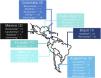
ResultsThe survey invitation response rate was 92.8% (n = 13/14 centers). Two institutions are university hospitals, and seven more correspond to centers where teaching and human resource training activities are carried out. On the other hand, only three institutions form part of the public hospital network of the region where they provide their services. The participating ECMO centers are located in seven Latin American countries (Fig. 1).

Only one center was not part of the ELSO. Among all the centers, a total of 133 devices were reported, the most widely used being Rotaflow® (23.3%, n = 31), followed by Centrimag® (20.3%, n = 27), Afinity® (15.8%, n = 21), Cardiohelp® (14.2%, n = 19), Revolution® (12.7%, n = 17), Biopump® (6.7%, n = 9), Bioconsole® (2.3%, n = 3) and Deltastream® (0.7%, n = 1). In addition, three centers reported having other devices (3.7%, n = 5) such as Stocker® (n = 3), and two other centers had Roller® (n = 2).
A total of 76.9% of the centers reported having mobile ECMO available (n = 10), with a total of 343 transfers being performed between 2011–2020, including 253 ground transfers and 90 air transfers.
A total of 1629 patients had undergone cannulation (342 in 2016; 292 in 2017; 325 in 2018; 250 in 2019; and 420 in 2020), with the majority being adults (62.5%, n = 1018; pediatric 28.7%, n = 468; newborn infants 8.8%, n = 143). In 2020, there were 218 suspected or confirmed COVID-19 patients receiving ECMO support, especially adults (98.6%, n = 215), with a much smaller number of pediatric (0.9%, n = 2) and neonatal (0.3%, n = 1) cases. These COVID-19 cases accounted for 51.9% of the total ECMO cases for that year, with a particular need for pulmonary ECMO (95.4%, n = 208) compared to cardiac ECMO (4.6%, n = 10).
Health professionals and assistant personnelThe centers reported a total of 310 specialist physicians as healthcare professionals, of which 70.3% (n = 218) had received ECMO training. The majority of the medical professionals were pediatric and adult critical care specialists (n = 75 and n = 71, respectively), of which 82.6% (n = 62) and 57.7% (n = 41) had ECMO training, respectively. The specialty with the largest number of ECMO-trained professionals was neonatology (97.4%, n = 37).
On the other hand, all centers reported having cardiovascular surgeons in their team (n = 52), and 80.7% (n = 42) had ECMO training, while 61.5% reported having anesthesiology professionals (n = 35), with 45.7% (n = 16) of them being trained in ECMO. Other reported specialties were thoracic surgery (n = 11, 54.5% ECMO specialists), pediatric surgery (n = 10, 60% ECMO specialists), internal medicine (n = 6, 50% ECMO specialists), and general surgery (n = 4, 25% ECMO specialists). Physicians specializing in pediatrics (n = 3), gynecology and obstetrics (n = 3), and emergency care medicine (n = 1) represented the least frequent specialties.
Some centers also reported having medical professionals specializing in toxicology, infectious diseases, cardio-pediatrics, hemodynamics, pulmonology, pediatric cardiovascular anesthesiology, intrathoracic transplant surgery, endovascular surgery and neurosurgery.
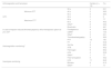
On the other hand, four of the 13 centers reported having general practitioners (n = 30) in their team, and up to 53.3% had ECMO training. In addition to medical professionals, other healthcare professionals such as nurses (n = 287, 87.1%; n = 250 with ECMO training) were reported. Other professionals included respiratory therapists (n = 55), perfusionists (n = 46), surgical instrument specialists (n = 31), physiotherapists (n = 19), speech therapists (n = 6), psychologists (n = 6) and occupational therapists (n = 5) (Table 1).
Other professions included in ECMO centers.
| Other professions | All | ECMO training | |
|---|---|---|---|
| n (%) | Yes | No | |
| n (%) | n (%) | ||
| General practitioners | 30 (6.2) | 16 (53.3) | 14 (46.7) |
| Nurses | 287 (59.2) | 250 (87.1) | 37 (12.9) |
| Perfusionists | 46 (9.5) | 45 (97.8) | 1 (2.2) |
| Surgical instrument specialists | 31 (6.4) | 17 (54.8) | 14 (45.2) |
| Respiratory therapists | 55 (11.3) | 32 (58.2) | 23 (41.8) |
| Physiotherapists | 19 (3.9) | 7 (36.8) | 12 (63.2) |
| Occupational therapists | 5 (1) | 0 (0) | 5 (100) |
| Phonoaudiologists | 6 (1.2) | 0 (0) | 6 (100) |
| Psychologists | 6 (1.2) | 1 (16.7) | 5 (83.3) |
In 53.8% of the centers, the direct patient care team consisted of an intensive care nurse in charge of the patient and a perfusionist or ECMO specialist in charge of the ECMO device. In the remaining centers, care was provided by a specialized ECMO nurse who was responsible for both the device and the patient. The nurse-to-patient ratio was 1:1 in most centers (76.9%, n = 10).
Performed interventionsThe most common indication for initiating ECMO support in adults was refractory hypoxemia (n = 310; cardiogenic shock n = 201, post-cardiotomy shock n = 89), whereas in pediatric patients the most common indication was post-cardiotomy shock (n = 174; cardiogenic shock n = 101, hypoxemia n = 55). Other indications were the postoperative period of heart or lung transplantation, protected angioplasty, diaphragmatic herniation and airway surgeries.
A total of 76.9% percent (n = 10) of the centers used some score for cannulation, with RESP being used in 69.2% (n = 9) of the centers, SAVE in 38.5% (n = 5) and PRESERVE in 30.8% (n = 4). The use of the Neo-RESCUERS and SOFA scores was also reported.
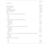
The most common ventilatory mode when starting ECMO was pressure control, with FiO2 ≤ 74% and PEEP 10−14 cm H2O. The most frequent programmed respiratory rate was 10−14 rpm, with a gas flow of 0−2 lpm especially in centers offering both pediatric and adult care (Table 2).
Management of mechanical ventilation.
| MV characteristics at the start of ECMO | Centers (n = 13) | (%) | |
|---|---|---|---|
| Ventilatory mode | Volume control | 6 | 46.2 |
| Pressure control | 7 | 53.8 | |
| FiO2 programming | <50% | 5 | 38.5 |
| 50−74% | 5 | 38.5 | |
| 75−90% | 0 | 0 | |
| >90% | 3 | 23.1 | |
| PEEP programming | 5−9 cmH2O | 4 | 30.8 |
| 10−14 cm H2O | 9 | 69.2 | |
| RR programming | 10−14 rpm | 11 | 84.6 |
| 15−19 rpm | 2 | 15.4 | |
| Gas flow programming in adult-only centers | 0−2 lpm | 2 | 33.3 |
| 3−5 lpm | 3 | 50.0 | |
| 6−8 lpm | 1 | 16.7 | |
| Gas flow programming in centers with care for adults and children | 0−2 lpm | 6 | 85.7 |
| 3−5 lpm | 1 | 14.3 | |
| 6−8 lpm | – | – | |
FIO2: fraction of inspired oxygen; PEEP: positive end-expiratory pressure. RR: respiratory rate.
Continuous RRT was the most commonly used form of renal replacement therapy, accounting for 76.9% of the cases. In 30.8% (n = 4) of the centers, RRT was directly connected to the patient.
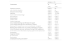
Concerning anticoagulation, unfractionated heparin (UFH) was used in all centers, with therapeutic ranges of minimum TTP of 30–50 s and a maximum of 40–60 s in most centers. The most common methods for monitoring anticoagulation were thromboplastin time, serum fibrinogen, and anti-factor Xa activity. LDH measurement was used to assess hemolysis in most centers. In cases of heparin-induced thrombocytopenia (HIT), the use of bivalirudin, fondaparinux, and/or low molecular weight heparin (LMWH) was reported (Table 3).
Anticoagulation in centers with ECMO therapy.
| Anticoagulation and hemolysis | Centers (n = 13) | (%) | ||
|---|---|---|---|---|
| UFH | Minimum PTT | 30 s | 3 | 23.1 |
| 40 s | 5 | 38.5 | ||
| 50 s | 5 | 38.5 | ||
| Maximum PTT | 40 s | 0 | 0 | |
| 50 s | 1 | 7.7 | ||
| 60 s | 6 | 46.2 | ||
| Otherb | 6 | 46.2 | ||
| In case of heparin-induced thrombocytopenia, which therapeutic option do you use? | Bivalirudin | 6 | 46.1 | |
| Fondaparinux | 4 | 30.8 | ||
| LMWH | 2 | 15.4 | ||
| Otherc | 3 | 23.1 | ||
| Anticoagulation monitoringa | Thromboelastography | 5 | 38.5 | |
| PTT | 13 | 100 | ||
| PT | 4 | 30.8 | ||
| Anti-Xa | 9 | 69.2 | ||
| ATIII | 6 | 46.2 | ||
| Fibrinogen | 9 | 69.2 | ||
| LDH | 4 | 30.8 | ||
| Otherd | 1 | 7.7 | ||
| Hemolysis monitoring | Free hemoglobin | 6 | 46.2 | |
| LDH | 11 | 84.6 | ||
| Bilirubin | 8 | 61.5 | ||
| Othere | 1 | 7.7 | ||
Circuit monitoring was most frequently performed by measuring pressures before and after the membrane in 92.3% (n = 12) of the centers, followed by temperature and extraction pressure. ICUs with ECMO that used circuit monitoring methods different from those contemplated in the survey indicated the use of venous return monitors.
To maintain hemostasis, packed red blood cell units were transfused with a hematocrit of 25% in most centers. In addition, anticoagulation was discontinued when the platelet count was under 30,000/mm3 or 50,000/mm3, and cryoprecipitates or fibrinogen concentrate were transfused when the fibrinogen level was 100 mg/dl. Transfusion of fresh frozen plasma was considered when the international normalized ratio (INR) was >2.0 in most centers (Table 4).
Characteristics of circuit monitoring and hemostasis without active bleeding.
| Characteristic | Centers |
|---|---|
| n = 13 | |
| n (%) | |
| Online O2 saturation | 8 (61.5) |
| Online hemoglobin | 7 (53.8) |
| Temperature | 11 (84.6) |
| Pre-membrane pressure | 12 (92.3) |
| Post-membrane pressure | 12 (92.3) |
| Extraction pressure | 10 (76.9) |
| Other | 2 (18.2) |
| Minimum hematocrit for transfusion of red blood cells | |
| 25% | 7 (53.8) |
| 30% | 3 (23.1) |
| 35% | 1 (7.7) |
| Other | 2 (15.4) |
| Minimum platelet count to discontinue anticoagulation | |
| <30.000/mm3 | 4 (30.8) |
| <40.000/mm3 | 3 (23.1) |
| <50.000/mm3 | 4 (30.8) |
| <60.000/mm3 | 1 (7.7) |
| Other | 1 (7.7)a |
| Minimum platelet count for transfusion of platelet concentrate | |
| <30.000/mm3 | 4 (30.8) |
| <40.000/mm3 | 1 (7.7) |
| <50.000/mm3 | 2 (15.4) |
| <60.000/mm3 | 3 (23.1) |
| Other | 3 (23.1) |
| Minimum fibrinogen value for transfusion of cryoprecipitates or fibrinogen concentrates | |
| <80 mg/dl | 1 (7.7) |
| 100 mg/dl | 9 (76.9) |
| 150 mg/dl | 3 (23.1) |
| Maximum INR for FFP transfusion | |
| >1.5 | – |
| >2.0 | 7 (53.8) |
| >2.5 | 4 (30.8) |
| >3.0 | 2 (15.4) |
INR: international normalized ratio, Hb: hemoglobin, FFP: fresh frozen plasma.
Patient perfusion monitoring was mainly performed at cerebral level (61.5%, n = 8), followed by peripheral monitoring (38.5%, n = 5), and one center (7.7%) reported performing renal NIRS monitoring. Sedation monitoring was performed using the RASS scale in 84.6% (n = 11) of the centers, and the NPASS scale in 15.4% (n = 2).
Regarding antibiotic prophylaxis, 5 centers (38.5%) administered antibiotics before cannulation, four centers (30.8%) administered antibiotics only in specific cases, and one center did not use antibiotic prophylaxis. In turn, prophylaxis during ECMO therapy without evidence of infection was performed in three centers (23.1%), while three other centers (23.1%) prescribed it on certain occasions without a protocol, and one center did not administer antibiotic prophylaxis.
OutcomesIn half of the centers, the length of stay was shorter in adults subjected to cardiac ECMO (50% 1–7 days, 33.3% 7–14 days, 16.7% no data available) than respiratory ECMO (15.4% 7–14 days, 53.8% 14–21 days, 23.1% >21 days, 7.7% ICU without ECMO availability for some patient type). On the other hand, in pediatric and neonatal patients, the duration of respiratory and cardiac ECMO therapy was less than 14 days (pediatric respiratory 46.1%, cardiac 53.8%; neonatal respiratory 30.7%, cardiac 53.8%).
The most frequent complications of V-A ECMO support were harlequin syndrome, left ventricular dilatation, intracranial hemorrhage and infections, followed by gastrointestinal tract bleeding. In the case of V-V ECMO support, infection was the most frequent complication, followed by circuit-related problems (recirculation, oxygenator failure and air in the circuit). Circuit-related bleeding requiring withdrawal of support was reported more frequently with V-A ECMO (Table 5).
Complications in Latin American ECMO centers according to type of ECMO.
| Complication | Center (n = 13) | |
|---|---|---|
| VA | VV | |
| n (%) | n (%) | |
| Harlequin syndrome | 9 (69.2) | NA |
| Left ventricular dilatation | 9 (69.2) | NA |
| Intracranial hemorrhage | 9 (69.2) | 6 (46.2) |
| Infections | 9 (69.2) | 10 (76.9) |
| Gastrointestinal hemorrhage | 8 (61.5) | 8 (61.5) |
| Pneumothorax | 4 (30.8) | 6 (46.2) |
| Decannulation | 3 (23.1) | 4 (30.8) |
| Cardiac arrest | 5 (38.5) | 5 (38.5) |
| Cardiac tamponade | 4 (30.8) | 0 (0.0) |
| Circuit-related bleeding and withdrawal of support | 5 (38.5) | 1 (7.7) |
| Circuit-related bleeding without withdrawal of support | 8 (61.5) | 8 (61.5) |
| Bleeding NOT related to the circuit and withdrawal of support | 6 (46.2) | 3 (23.1) |
| Bleeding NOT related to the circuit without withdrawal of support | 8 (61.5) | 8 (61.5) |
| Circuit thrombosis (cannula, membrane) forcing ECMO suspension. | 3 (23.1) | 6 (46.2) |
| Acute massive circuit thrombosis requiring urgent circuit replacement | 3 (23.1) | 4 (30.8) |
| Pump failure | 1 (7.7) | 2 (15.4) |
| Oxygenator failure | 5 (38.5) | 9 (69.2) |
| Air in the circuit | 7 (53.8) | 9 (69.2) |
| Recirculation | NA | 9 (69.2) |
| Complications during cannulationa | 2 (16.7) | 4 (30.8) |
NA: not applicable.
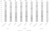
The overall survival rate at the time of decannulation was 55.7% (95%CI 53.0–58.3). In the pediatric group, the highest survival rate corresponded to cardiac ECMO, while in the adult and neonatal population, the highest survival rate corresponded to respiratory ECMO (Fig. 2).
Discussion
The present study describes the general characteristics of different ECMO centers located throughout Latin America. The participating ICUs with ECMO service were mostly university and teaching hospitals, with only a few of them belonging to the regional public network. A diversity of ECMO devices was reported, with Rotaflow® and Centrimag® being the most common. Most centers have mobile ECMO and treated various types of patients, with a notable increase in COVID-19 cases. ECMO training is widespread among professionals, and overall survival at decannulation varies according to the type of ECMO support provided.
According to the ELSO registry, between 2016 and 2020 there were 567 member centers worldwide, reporting a total of 77,922 cannulations during that period. Of these centers, 46 were in Latin America (n = 28 ICUs with ECMO service in 2016, and n = 46 in 2020), with 2074 cannulations performed during that period.11 The centers participating in this study recorded approximately 300 patients per year receiving ECMO therapy support—a figure that increased to more than 400 patients by 2020. This increase was especially notable for respiratory support in adults with acute respiratory distress syndrome (ARDS) secondary to COVID-19. On the other hand, some European centers reported an increase in the number of cannulations, especially in patients with cardiogenic shock who required V-A ECMO support between 2010 and 2015.12 The number of ECMO patients in the participating institutions doubled the number reported in cohort studies conducted at 60 hospitals in the United States between March and July 2020.13 Furthermore, the number was significantly higher than the figures reported in single-center studies conducted in different Latin American countries.14 However, in addition to the number of patients receiving ECMO support, which reflects the care capacity acquired by the institution, it is essential to emphasize the reported outcomes. For ELSO centers in Latin America between 2018 and 2022, the adult patient survival rates up to decannulation or transfer in pulmonary ECMO (54%), cardiac ECMO (53%) and extracorporeal cardiopulmonary resuscitation (ECPR) (32%)11 were lower compared to the survival rates reported in our study. In this context, when comparing the survival outcomes with other studies found in the literature, we observed that the survival rate among adults with respiratory ECMO support was lower in our study than in the United Kingdom (74.0%)15 in a retrospective cohort of 1205 patients between 2011 and 2017. However, the survival rate was higher than that reported in a nationwide analysis in Germany conducted between 2007 and 2018.16 In pediatric cases, survival data recorded by the ELSO were consistent with our own findings (74% in pulmonary ECMO, 61% in cardiac ECMO, and 34% in ECPR).11 Other studies in the pediatric and neonatal population have also recorded survival rates of 74% at disconnection and 57% at hospital discharge, with no significant differences according to the type of ECMO involved.17
The training of healthcare professionals who assist ECMO patients is a crucial aspect for both effective functioning of the center and positive patient outcomes. We recorded specialists in various areas such as high complexity surgery, intensive and perioperative care, perinatal maternal care, and emergency patient care. In addition, the centers have dedicated personnel in charge of providing ECMO support, adapting intervention to the natural progression of each case, with the ultimate goal of securing comprehensive recovery and rehabilitation. These findings are in line with the existing literature, which emphasizes that factors associated with high-quality ECMO programs include ongoing education and training for personnel, state-of-the-art facilities and technology, and a well-organized and experienced ECMO center. In addition, these factors are closely linked to the number of cases treated annually.18–20
Regarding the ventilation practices employed in the participating ICUs, they consider the use of pressure control, followed by volume control. In this regard, according to an international survey conducted in 141 centers in 28 countries, only 27% of the centers had a specific protocol for mechanical ventilation in patients undergoing ECMO therapy, the main objective of ventilation being "lung rest". In addition, 76% of the respondents aimed for a target tidal volume of6 ml/kg or less, applied positive end-expiratory pressure, and set a positive pressure of 6–10 cm H2O when ventilating patients with V-V ECMO.21
ECMO support in critically ill patients, as a widely implemented intervention worldwide that moreover increased during the COVID-19 pandemic, faces substantial challenges in our region. A notable strength identified in this study is the high response rate to the invitation to participate. Although the invitation was not extended to all existing units in the region but to those that formed part of a closed group created through social networks, the participating ICUs with ECMO service were distributed throughout Latin America, offering indirect knowledge about the practices implemented in different countries. According to the current literature, ECMOsupport has increased greatly in the last two decades. In this regard, the COVID-19 pandemic was a period that allowed the opening of new ECMO centers, and at the same time consolidated the technique in those centers that were already using it. As a result, the present study does not include centers that have recently become operational, as well as some centers with experience that did not participate in the survey. Nevertheless, this initial survey reflects efforts to adhere to and maintain the ELSO standards for appropriate patient care. In addition, based on the survival results, the participating centers were able to provide care comparable to that afforded by ECMO units in high-income countries.
Given the retrospective nature of this study, the availability of data referred to certain variables of interest may be limited, including the criteria for starting ECMO support. Although there are clinical criteria that help define the initiation of extracorporeal circulatory support, including patient weight, days on mechanical ventilation, ventilatory mode and many more, these criteria vary according to the different intensive care groups and the clinical condition of the patient. It thus would be important for future reports to include aspects which, in the light of current evidence, are considered to represent good clinical practice, such as low tidal volumes, neuromuscular blocking agents and prone positioning, among others. Therefore, continued efforts should focus on maintaining adequate and standardized registers of centers and patients, allowing comparisons with other ECMO units.
ECMO therapy is a complex and high-risk treatment indicated in patients with the most severe forms of acute respiratory failure and cardiogenic shock, as well as in highly complex cardiovascular surgery scenarios. Despite the intrinsic risks associated with ECMO therapy, morbidity and mortality can be reduced in centers with specific management protocols and strict patient selection criteria.22,23
ConclusionsBetween 2016 and 2020, the ICUs with ECMO service participating in this study and located in Latin America have taken care of critically ill adult, pediatric and neonatal patients. Such care has been provided in an organized manner, allowing similar survival outcomes to those obtained in other ECMO units (regionally and in other continents). This allows us to compare ourselves with the international standards referred by the ELSO. It is necessary to strengthen a communication and information network at the Latin American level to collect data on ECMO practices, which will facilitate scientific and academic support among the different groups, thus promoting the use of the technique in the region. This initiative aims to generate greater interest in the reporting and publication of ECMO cases, which will increase the visibility of the region and provide clinical evidence on its care practice. Future initiatives such as this will seek to generate more interest and allow for subgroup analyses according to models of care or types of ECMO support applications.
Latin America is making progress in the implementation of ECMO therapy with ELSO support, which facilitates the generation of local information and the comparison of global results. However, it is crucial to continue optimizing data recording to improve the monitoring and evaluation of treatments. This study represents a preliminary effort to gather evidence on the structural and functional capabilities of ECMO units in Latin America.
CRediT authorship contribution statementCamilo Pizarro: conception and design of the study, data acquisition, analysis and interpretation of the data, revision of the draft article and critical review of the intellectual content, final approval.
Anderson Bermon: conception and design of the study, data acquisition, analysis and interpretation of the data, revision of the draft article and critical review of the intellectual content, final approval.
Silvia Plata Vanegas: conception and design of the study, data acquisition, analysis and interpretation of the data, revision of the draft article and critical review of the intellectual content, final approval.
Claudia Colmenares-Mejia: conception and design of the study, data acquisition, analysis and interpretation of the data, revision of the draft article and critical review of the intellectual content, final approval.
Claudia Marcela Poveda: data acquisition, analysis and interpretation of the data, revision of the draft article and critical review of the intellectual content, final approval.
René D. Gómez Gutiérrez: data acquisition, analysis and interpretation of the data, revision of the draft article and critical review of the intellectual content, final approval.
Jorge Arturo Ramírez Arce: data acquisition, analysis and interpretation of the data, revision of the draft article and critical review of the intellectual content, final approval.
Sonia Villarroel: data acquisition, analysis and interpretation of the data, revision of the draft article and critical review of the intellectual content, final approval.
Daniel Absi: data acquisition, analysis and interpretation of the data, revision of the draft article and critical review of the intellectual content, final approval.
Marco Antonio Montes de Oca Sandoval: data acquisition, analysis and interpretation of the data, revision of the article draft and critical review of the intellectual content, final approval.
Fernando Pálizas: data acquisition, analysis and interpretation of the data, revision of the article draft and critical review of the intellectual content, final approval.
Leonardo Salazar: conception and design of the study, review of the draft article and critical review of the intellectual content.
FundingThis article received no funding from internal or external institutions.
Special thanks to Mary Alejandra Mendoza (Fundación Cardiovascular de Colombia), Mariano Norese, Christian Casabella García, Fernando Pálizas (Clínica Bazterrica), María Luisa Pilan, Gisela Ponce (Hospital de Pediatría J.P. Garrahan), Luiz Fernando Caneo (InCor-HC-FMUSP), Silvio Fabio Torres (Hospital Austral), Vadim Kotowicz, Luisa Baldini, Mateo Ferrero (Hospital Italiano), Telmo Fernández Cadena (Hospital Luis Vernaza. Guayaquil, Ecuador), Jorge Rufs (Clínica Las Condes) and Walter Mogrovejo Ramos, Moisés Vidal Lostaunau (Clínica del INCA) for their support and participation in this study.

