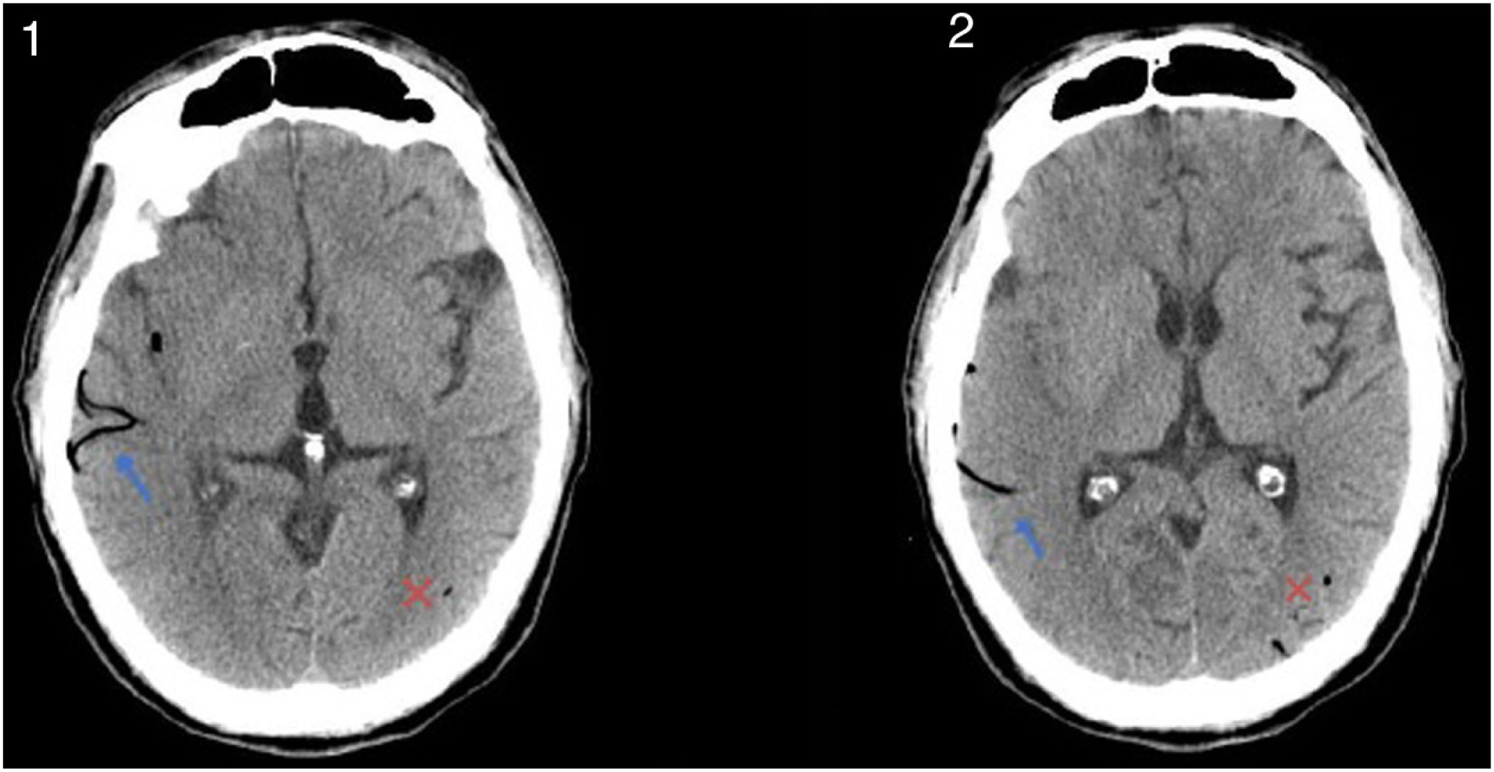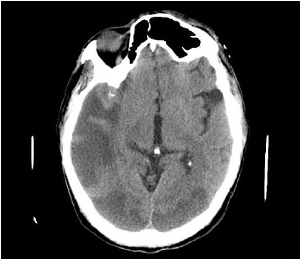
This is the case of a 47-yearl old man with a past medical history of native mitral aortic valve acute endocarditis who required valvular repair and supracoronary tube implantation. The patient is admitted to the ICU after a scheduled reintervention due to severe paravalvular leak followed by valvular replacement with mechanical prosthesis. Initially extubated and without neurologic focality. A few minutes after experiencing the early seizure the patient requires orotracheal intubation. The emergency cranial CT scan performed (Fig. 1) reveals the presence of air into the right middle cerebral artery territory, M2 (blue arrow [the color of the figure can only be seen on the electronic version]) and part of its cortical branches, as well as air bubbles into the left posterior cerebral artery (x) that consistent with gas embolism. The radiographic follow-up performed 48 h later (Fig. 2) reveals the presence of an extensive established right parietal-temporal-occipital and left occipital ischemic infarction with slight midline deviation.
Impact factor
The Impact Factor measures the average number of citations received in a particular year by papers published in the journal during the two preceding years.
© Clarivate Analytics, Journal Citation Reports 2025
Impact factor 2024
3.1
Citescore 2024
2.3
SJR
SRJ is a prestige metric based on the idea that not all citations are the same. SJR uses a similar algorithm as the Google page rank; it provides a quantitative and qualitative measure of the journal's impact.
See moreSJR 2024
0.353
SNIP
SNIP measures contextual citation impact by wighting citations based on the total number of citations in a subject field.
See moreSNIP 2024
0.573
View more metrics