


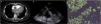
A 61-year-old woman with a history of liver cirrhosis presented to the hospital with a one-week history of chest tightness and dizziness. Computed tomographic pulmonary angiography revealed a large-area filling defect in the right atrium (Fig. 1 Panel A). Transesophageal echocardiography was then performed and found a giant mass almost occupying the entire atrial cavity (Fig. 1 Panel B, Video 1), and color Doppler showed a small amount of blood flow passed through the gap between the mass and the atrial wall (Video 2). Subsequently, the patient underwent a thoracotomy and planned to receive a scheduled resection of the atrial mass. During thoracotomy, it was observed that the thymus, aortic wall, and pulmonary artery wall were extensively invaded. The patient's relatives finally refused to undergo further atrial mass resection given the expected high risk of perioperative death. Thymic pathology and immunohistochemical results indicated diffuse large B-cell lymphoma (Fig. 1 Panel C). Based on these findings, cardiac diffuse large B-cell lymphoma was highly suspected, which is a rare and life-threatening clinical condition.
Contribution of the authors
Dong P drafted the manuscript and revised the manuscript. Pan J participated in the patient's care and drafted the manuscript. Zhou X participated in the patient's care and revised the manuscript. All authors read and approved the final manuscript.
FundingThis work was supported by grants from the Zhejiang Medicine and Health Science and Technology Project (No. 2023KY1084) and the Project of NINGBO Leading Medical & Health Discipline (No. 2022-F16). The funders had no role in the study design, data collection, and analysis, decision to publish, or manuscript preparation.
Conflicts of interestThe authors declare that they have no potential conflict of interest.
Consent for publicationWritten informed consent was obtained from the patient for publication of this article and any accompanying images.
None.

