


A 70-year-old male reported to the emergency service due to recurrent abdominal pain associated with physical exertion. He tended to have low blood pressure, and the physical examination revealed a pulsatile mesogastric mass measuring about 10 cm in diameter.
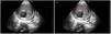
Point-of-care ultrasound (POCUS) revealed an infrarenal aortic aneurysm measuring 11 cm in diameter and with an internal lumen of 4 cm (Fig. 1) ventral to the hyperechogenic zone with a posterior shadow corresponding to the spine (V). The characteristic signs of a thrombosed aortic aneurysm can be observed, with a turbulent internal flow (“swirling smoke” pattern), surrounded by the thrombosed zone measuring up to 7 cm in thickness (T). The accompanying video evidences the pulsatility of the aorta and the turbulent flow within the lumen (Video).
Author’s contributions
All three authors, as a working team, have contributed similarly to the diagnosis and treatment of the patient, and in the preparation and writing of the manuscript.
FundingThis study has received no funding.

