


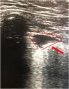

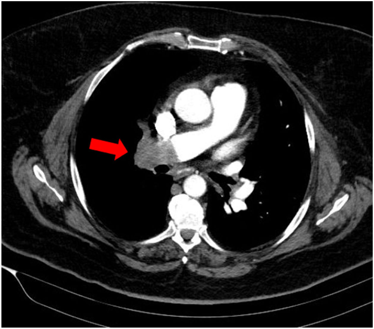
This is the case of a 61-year-old woman with obesity, hypertension, and chronic obstructive pulmonary disease (COPD) who presented to the emergency room with clinical signs of dyspnea and pleuritic pain in right hemothorax of 10-day evolution. She showed significant respiratory effort, which is why non-invasive mechanical ventilation (NIMV) was started. Thoracic X-ray looked normal, and the electrocardiogram performed revealed the presence of a dilated—though normocontractile—right ventricle. The thoracic ultrasound revealed the presence of a triangular hypoechogenic image of peripheral base that was consistent with right pulmonary infarction (Fig. 1). The axial thoracic computed tomography (CT) scan showed a repletion defect in the right pulmonary artery spreading towards the lobar branches plus pulmonary alveolar damage to the right upper lobe suggestive of associated pulmonary infarction (Fig. 2). The ultrasound provides extremely valuable additional information—essential at times—for the optimal management of the patients. The most indicative finding on the thoracic ultrasound of pulmonary thromboembolism (PTE) is the presence of triangular hypoechoic lesions and pleural base surrounded by a pleural line of normal aspect with or without associated pleural effusion.
Funding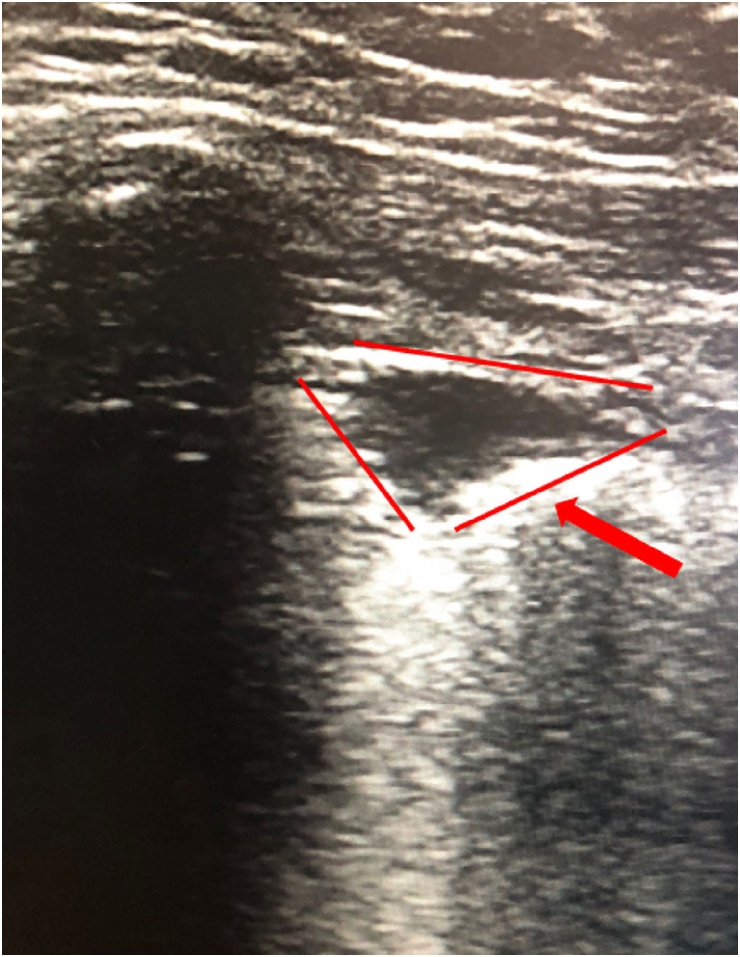
None whatsoever.
We wish to thank the intensive care unit of Hospital Clínico Lozano Blesa, Zaragoza, Spain.
Please cite this article as: Edroso Jarne PE, Matute Guerrero A, Vicho Pereira R. Diagnóstico de infarto pulmonar por ecografía torácica en paciente con tromboembolismo pulmonar masivo. Med Intensiva. 2022;46:601–602.

