


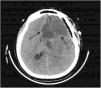
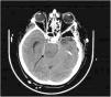
This is the case of a 75-year-old man treated with scheduled surgical clipping of a giant aneurysm at left internal carotid artery level. After surgery, a cranial CT scan performed revealed the presence of an image consistent with acute ischemia in the left frontal region. Twenty-four hours later, right side mydriasis was reported. This new finding prompted a new cranial CT scan that revealed the progression of ischemic phenomena with damage to the left middle and posterior cerebral artery territories (Fig. 1) with signs of intracranial hypertension and compression at brainstem level against the contralateral tentorium (Fig. 2), which would explain the midriasis of the right side. This is know as the Kernohan-Woltman notch phenomenon. Despite medical treatment, the option of decompressive craniectomy was ruled out, and the patient progressed to brain death.
El factor de impacto mide la media del número de citaciones recibidas en un año por trabajos publicados en la publicación durante los dos años anteriores.
© Clarivate Analytics, Journal Citation Reports 2025
SJR es una prestigiosa métrica basada en la idea de que todas las citaciones no son iguales. SJR usa un algoritmo similar al page rank de Google; es una medida cuantitativa y cualitativa al impacto de una publicación.
Ver másSNIP permite comparar el impacto de revistas de diferentes campos temáticos, corrigiendo las diferencias en la probabilidad de ser citado que existe entre revistas de distintas materias.
Ver más


