


A 49-year-old woman with a history of bariatric surgery and liver cirrhosis was admitted due to upper gastrointestinal bleeding. Endoscopy evidenced 5 large esophageal varicose strands, with abundant bleeding. Ligation with elastic bands was attempted but proved unsuccessful, and a self-expanding stent had to be placed.
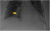
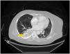
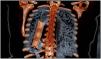
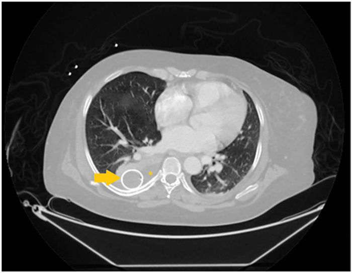
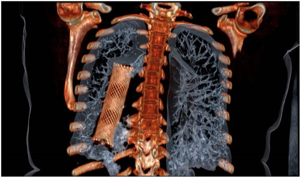
Radiological control (Fig. 1) and thoracoabdominal computed tomography angiography (CTA) (Figs. 2 and 3) identified a foreign body consistent with the stent and located in the right pleural space, with pleural effusion associated with compressive atelectasis and no evidence of esophageal wall damage. The situation was evaluated by Thoracic Surgery, and the stent was extracted without incidents via a mini-thoracotomy.
Declaration of Generative AI and AI-assisted technologies in the writing process
No artificial intelligence tool was used in the preparation of this work.
Financial supportThe present study has received no financial support.
Conflicts of interestThe authors declare that they have no conflicts of interest.

