


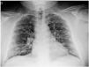


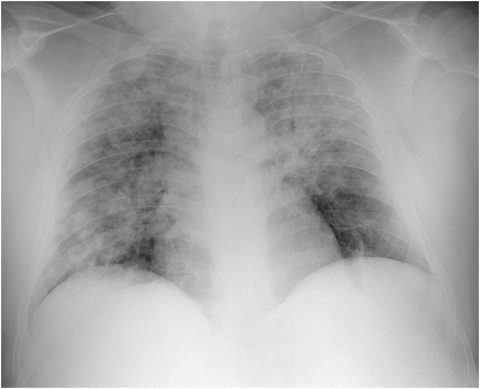
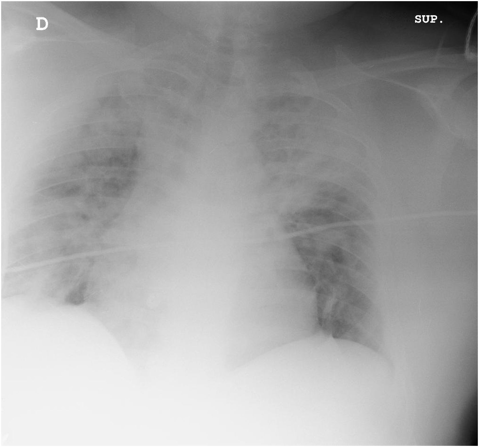
This is the case of a 51-year-old male with metabolic syndrome, chronic nephropathy, scleredema diabeticorum, and obstructive sleep apnea syndrome; he is admitted to the emergency room with 100.4 °F fever, dyspnea, and 5-day evolution green sputum. Blood cultures are drawn, and Legionella and pneumococcus urine antigens tests are performed together with several respiratory viral panel tests (RSV, Influenza, rhinovirus). A thoracic X-ray is performed (Fig. 1). The patient is admitted to the hospital, and wide-spectrum antibiotic therapy and oxygen therapy are administered due to hypoxemia (O2 saturation < 90%). Due to progressive worsening, both clinical—requiring high-flow nasal cannula—and radiologic (Fig. 2) admission to the Intensive Care Unit is decided (Fig. 3). Due to the patient’s torpid progression, and lack of microbiological isolates, a nasopharyngeal PCR swab testing is performed for SARS-CoV2 that tests positive.

Please cite this article as: Álvarez Méndez A, Matía Almudévar P, Álvarez Hernández S. Neumonía bilateral por SARS-CoV2: evolución radiográfica. Med Intensiva. 2022;46:420–421.

