


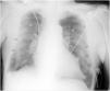
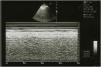
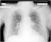
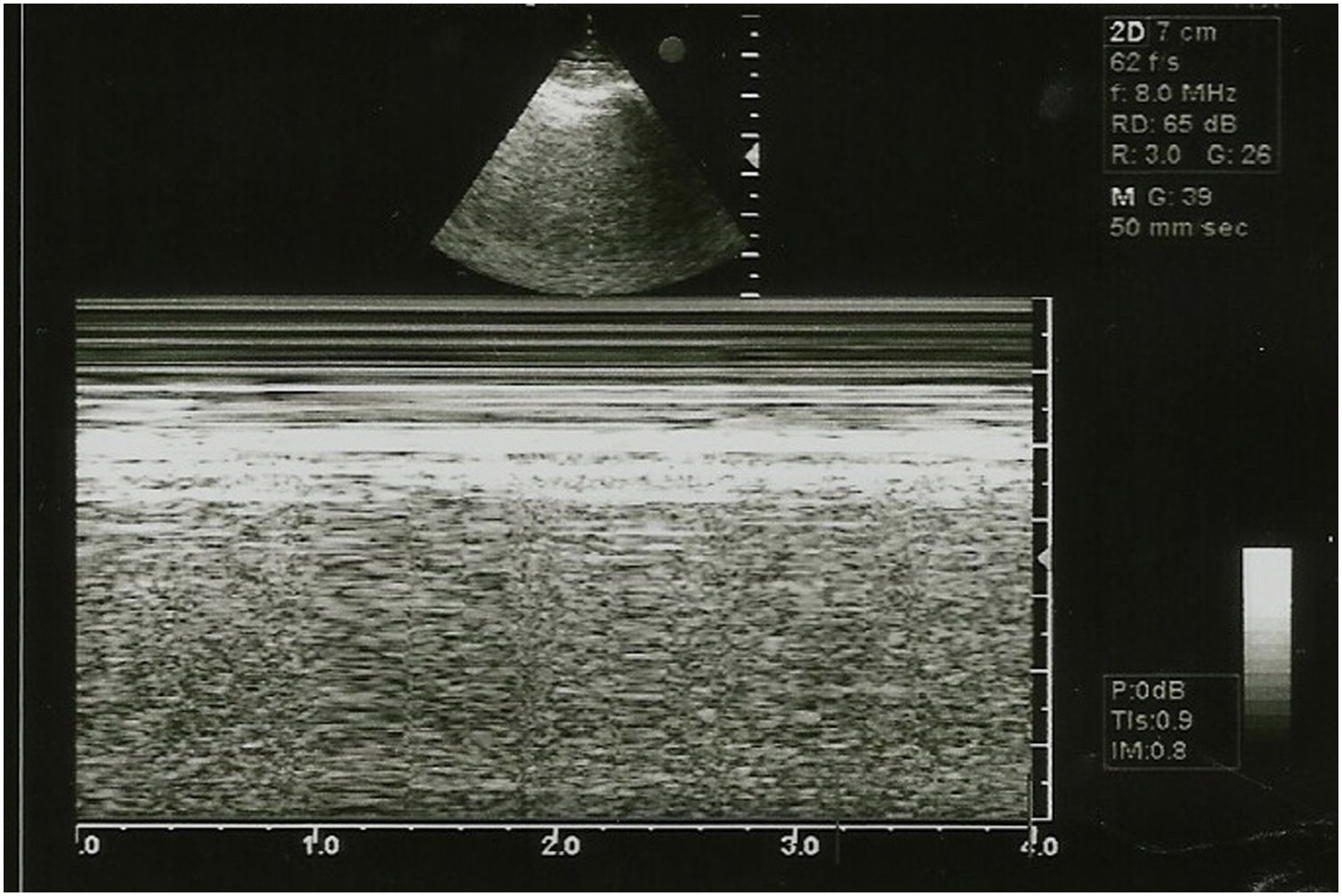
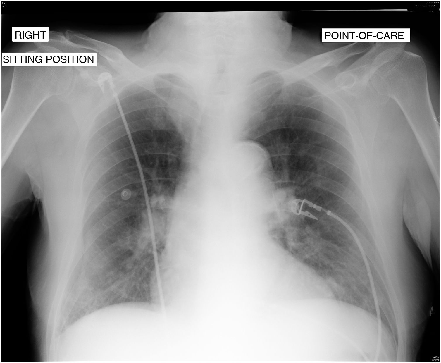
This is the case of an 86-year-old man admitted to our unit after emergency percutaneous coronary intervention due to anterior ST-segment elevation acute myocardial infarction with implantation of 2 drug-eluting stents into the LAD and first diagonal branch. Upon admission the situation of the patient remains hemodynamically and respiratory stable with 93% oxygen saturation on pulse oximetry without oxygen supply. A thoracic x-ray (Rx) is performed in the supine position that reveals the presence of 2 lines suggestive of bilateral pneumothorax (Fig. 1, arrows) that cannot be seen in the upper lobes (Fig. 1, arrowheads). To rule out the presence of bilateral pneumothorax, a bilateral thoracic ultrasound is performed that reveals the presence of pleural «sliding» on the imaging in M-mode or «seashore sign» (Fig. 2). In an Rx performed afterwards while in the semi-sitting position, only a small right baseline line can be seen (Fig. 3), and diagnosis of false bilateral skin fold pneumothorax is achieved.
Please cite this article as: Monterrubio Villar J, Almaraz Velarde R. Falso neumotórax bilateral secundario a pliegue cutáneo: utilidad de la ecografía torácica para su diagnóstico. Med Intensiva. 2022;46:357–358.

