
Edited by: Juan Antonio Llompart-Pou - Specialist in Intensive Care Medicine, Doctorate in Health Sciences, Universitat Illes Balears. Department of Intensive Care Medicine, Trauma and Neurocritical ICU, Hospital Universitari Son Espases. Palma, Spain
Last update: April 2026
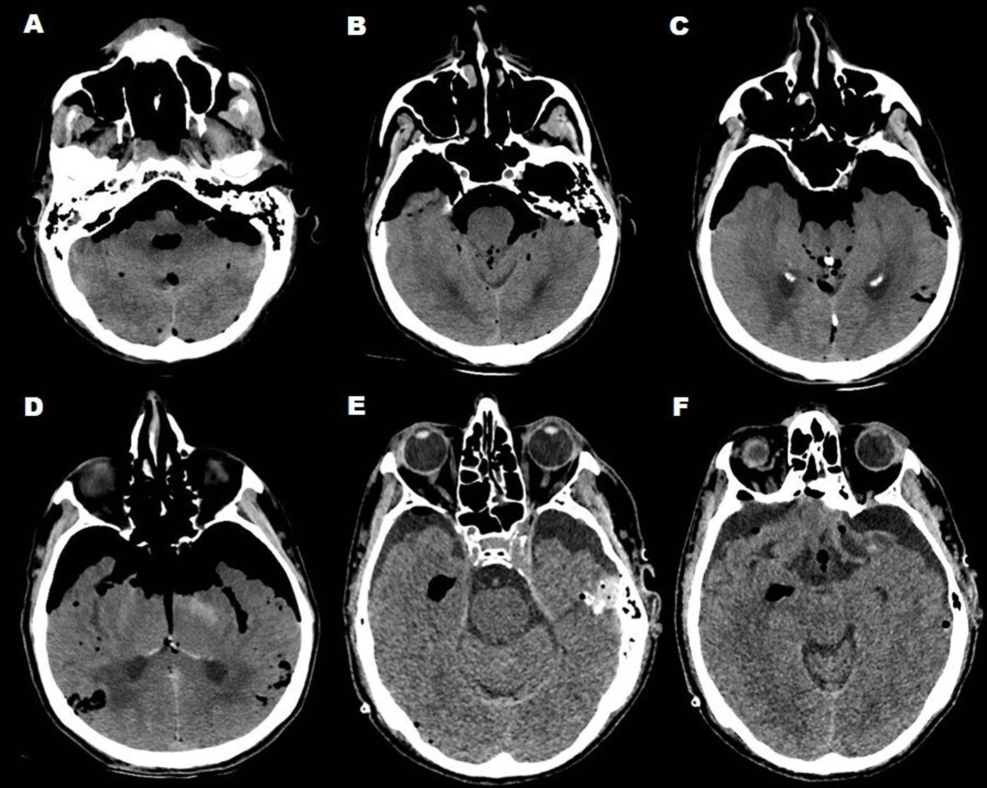
More infoA 77-year-old man presents with a traumatic brain injury (fall from 2 m) and progressive deterioration of consciousness. He arrives at the emergency department with a GCS of 3/15 and right-side otorrhagia. He is admitted to the intensive care unit on mechanical ventilation, without motor response, and with preserved brainstem reflexes. Brain CT shows extensive pneumocephalus involving the posterior fossa and bifrontal regions (Fig. 1A–D), as well as a left gangliobasal hemorrhage (Fig. 1D). Emergency neurosurgical decompression is performed. The patient shows postoperative improvement, with eye opening to voice and motor localization responses. Follow-up computed tomography demonstrated a decrease in the pneumocephalus (Fig. 1E–F). Tension pneumocephalus is a neurosurgical emergency that requires prompt diagnosis and treatment due to its imminent risk. Mild cases require 100% supplemental oxygen, but when neurological deterioration or mass effect is present, surgery is necessary.
None declared.