
Suggestions
Idioma
Guide for authors
Searcher
Journal Information
Cite
Cite
Share
Download PDF
More article options
Visits
1111
Scientific Letter
Full text access

High serum malondialdehyde levels in patients with decompression sickness

Altos niveles séricos de malondialdehído en pacientes con enfermedad descompresiva
Visits
1111
Leonardo Lorentea,
Corresponding author
lorentemartin@msn.com

Corresponding author.
, Pedro Abreu Gonzálezb, Candelaria Darias Martina, Ana Matutea, Gabriela Menéndeza, Juan Francisco Lozano Gomariza, Alejandro Jiménezc
a Intensive Care Unit, Hospital Universitario de Canarias, Ofra s/n. San Cristóbal de La Laguna, 38320, Tenerife, Spain
b Department of Physiology, Faculty of Medicine, University of La Laguna, Ofra, s/n. San Cristóbal de La Laguna, 38320, Tenerife, Spain
c Research Unit, Hospital Universitario de Canarias, Ofra, s/n. La Laguna, 38320, Santa Cruz de Tenerife, Spain
This item has received
Article information
Full Text
Bibliography
Download PDF
Statistics
Tables (2)
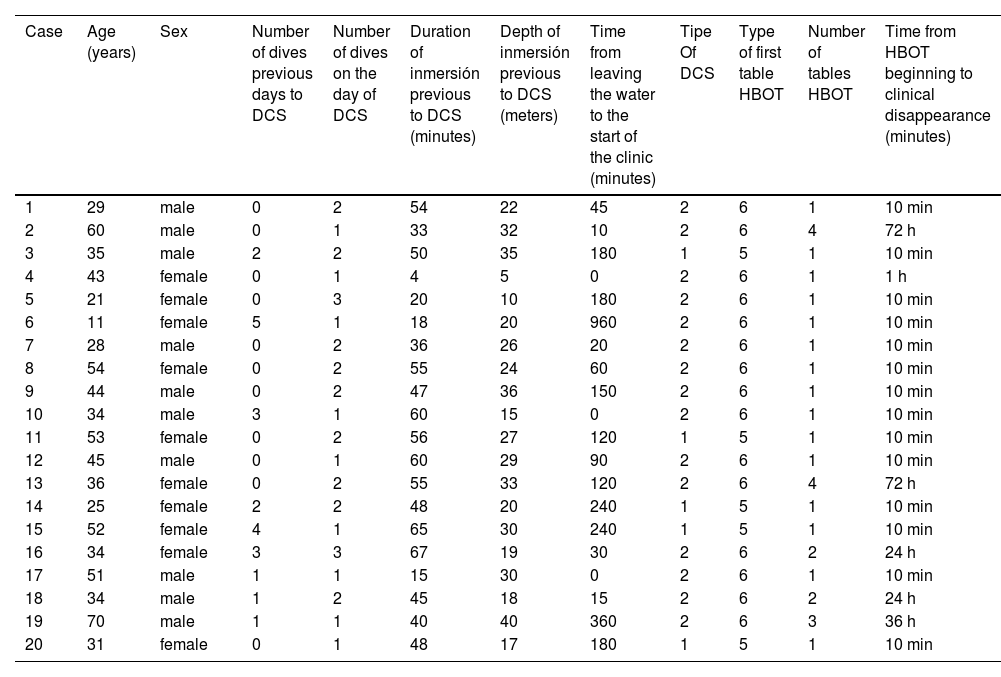
Table 1. Clinical characteristics of patients with decompression sickness (DCS).
Tables
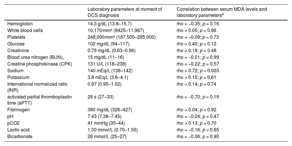
Table 2. Laboratory parameters at moment of decompression sickness (DCS) diagnosis and its association with serum malondialdehyde (MDA) levels.
Tables
Glossary
Full Text

The primary injury mechanism in decompression sickness (DCS) is the formation of bubble during dives, and it is widely accepted that its definitive treatment consists of hyperbaric oxygen therapy (HBOT) to reduce bubble volume.1

In addition, some published data have reported an increase in oxidative stress in DCS2–4 and that HBOT may influence oxidative stress.2–7 In two previous studies with rats, it was found that DCS increases malondialdehyde (MDA), a lipid hyperoxidation biomarker,8 in spinal cord and brain biopsies, and that the HBOT reduces those levels.2,3 One study reported a subject with DCS who underwent HBOT and showed an increase in salivary reactive oxygen species (ROS), urinary nitric oxide metabolites and urinary 8-isoprostane (a lipid peroxidation biomarker), all of which increased further after HBOT.4 In some studies, with healthy volunteers who underwent HBOT was found after HBOT an increase of ROS and glutathione peroxidase (antioxidant) in lymphocytes,5 of salivary ROS, of urinary 8-isoprostane, of salivary total antioxidant capacity (TAC)6 and of serum superoxide dismutase (antioxidant) and of serum MDA.7 In addition, an increase in oxidative stress has been reported in individuals who practice diving with self-contained underwater breathing apparatus (scuba).4,5,9,10 Therefore, we think that serum MDA levels could be increased in DCS patients.

The novel objectives of our study were to compare serum MDA levels between DCS patients and healthy subjects, and to determine the effect of HBOT on those serum MDA levels in DCS patients.

This prospective observational study was conducted with the approval of the Clinical Research Ethics Committee of the Hospital Universitario de Canarias (HUC), Tenerife, Spain (code CHUC_2023_150; 21st March 2024) and with informed consent signed by scuba divers with DCS and healthy subjects. Patient recruitment was carried out in the hyperbaric section of the Tenerife Medical Institute Limited Company (IMETISA), and its assistance activity is carried out for staff of Intensive Medicine Service of HUC.

Serum samples were obtained from DCS patients (at the time of diagnosis and 12 h after undergoing HBOT) and healthy subjects. We obtained the second blood sample after 12 h of undergoing HBOT because we had observed that most DCS patients needed only one HBOT session and lived far from the hyperbaric chamber; thus, it would have been difficult to obtain a second sample later. Serum MDA concentrations were determined using the thiobarbituric acid-reactive substances (TBARS) method previously described by Kikugawa et al.8

DCS patients received HBOT in a hyperbaric chamber, model IBERCO IB-230 × 6000. We administered HBOT following U.S. Navy recommendations.1 HBOT table 5 was used for the treatment of type I DCS, which involves musculoskeletal pain and skin symptoms, and the patient remains at 2.8 atmospheres absolute (ATA) or 18 m of seawater (msw) or 60 feet of seawater (fsw) for 45 min (40 min with 100% oxygen and 5 min with air at 21% oxygen). HBOT table 6 was used for the treatment of type II DCS, which involves the central nervous system, the inner ear, or the cardiopulmonary system; and the patient remains at 2.8 ATA for 75 min (60 min with 100% oxygen and 15 min with air at 21% oxygen).

There were no significant differences between DCS patients (n = 20) and healthy subjects (n = 17) in age (36 (30–52) vs 36 (28–49) years; p = 0.75) and sex (10 (50%) vs 8 (47%; p = 0.99). We found lower serum MDA levels in healthy controls (0.68 [0.61−0.74] nmol/mL) than in DCS patients at the time of diagnosis (1.38 (1.04–2.12) nmol/mL); p < 0.001) and after first HBOT (1.32 (1.18–1.85) nmol/mL); p < 0.001; n = 17). However, there were no significant differences in serum MDA levels in DCS patients at moment of DCS diagnosis and after first HBOT (p = 0.69).

The characteristics of dives and of HBOT sessions in each DCS patient are shown in Table 1. There were no significant associations between serum MDA levels at the time of DCS diagnosis and type of DCS (p = 0.15), sex (p = 0.40), age (rho = 0.16; p = 0.49), number of dives in previous days (rho = 0.09; p = 0.70), number of dives on the day of the event (rho = 0.32; p = 0.16), time of last immersion (rho = 0.57; p = 0.01), depth of last immersion (rho = 0.13; p = 0.58), time from leaving the water to symptom onset (rho= −0.17; p = 0.48), number of sessions of HBOT (rho = 0.14; p = 0.57), time from HBOT initiation to symptom resolution (rho = 0.06; p = 0.81).

Table 1.

Clinical characteristics of patients with decompression sickness (DCS).

Case  Age (years)  Sex  Number of dives previous days to DCS  Number of dives on the day of DCS  Duration of inmersión previous to DCS (minutes)  Depth of inmersión previous to DCS (meters)  Time from leaving the water to the start of the clinic (minutes)  Tipe Of DCS  Type of first table HBOT  Number of tables HBOT  Time from HBOT beginning to clinical disappearance (minutes) 
29  male  54  22  45  10 min 
60  male  33  32  10  72 h 
35  male  50  35  180  10 min 
43  female  1 h 
21  female  20  10  180  10 min 
11  female  18  20  960  10 min 
28  male  36  26  20  10 min 
54  female  55  24  60  10 min 
44  male  47  36  150  10 min 
10  34  male  60  15  10 min 
11  53  female  56  27  120  10 min 
12  45  male  60  29  90  10 min 
13  36  female  55  33  120  72 h 
14  25  female  48  20  240  10 min 
15  52  female  65  30  240  10 min 
16  34  female  67  19  30  24 h 
17  51  male  15  30  10 min 
18  34  male  45  18  15  24 h 
19  70  male  40  40  360  36 h 
20  31  female  48  17  180  10 min 

HBOT: hyperbaric oxygen treatment.

There were no significant associations between laboratory parameters and serum MDA levels at moment of DCS diagnosis (Table 2). No DCS patient needed mechanical ventilation, vasoactive agents, or Intensive Care Unit stay.

Table 2.

Laboratory parameters at moment of decompression sickness (DCS) diagnosis and its association with serum malondialdehyde (MDA) levels.

  Laboratory parameters at moment of DCS diagnosis  Correlation between serum MDA levels and laboratory parametersa 
Hemoglobin  14.3 g/dL (13.8–15.7)  rho = −0.35; p = 0.16 
White blood cells  10,170/mm³ (9425–11,987)  rho = 0.05; p = 0.86 
Platelets  248,000/mm³ (187,500–285 000)  rho = −0.09; p = 0.73 
Glucose  102 mg/dL (94–117)  rho = 0.40; p = 0.12 
Creatinine  0.79 mg/dL (0.63–0.96)  rho = 0.18; p = 0.48 
Blood urea nitrogen (BUN),  15 mg/dL (11–16)  rho = −0.01; p = 0.99 
Creatine phosphokinase (CPK)  131 U/L (118–238)  rho = −0.22; p = 0.57 
Sodium  140 mEq/L (138–142)  rho = 0.72; p = 0.003 
Potassium  3.8 mEq/L (3.6–4.1)  rho = 0.15; p = 0.61 
International normalized ratio (INR)  0.97 (0.90–1.02)  rho = 0.14; p = 0.74 
activated partial thromboplastin time (aPTT)  28 s (27–33)  rho = −0.70; p = 0.19 
Fibrinogen  380 mg/dL (326–427)  rho = 0.04; p = 0.92 
pH  7.43 (7.38–7.45)  rho = −0.24; p = 0.47 
pCO2  41 mmHg (35–44)  rho = 0.13; p = 0.70 
Lactic acid  1.00 mmol/L (0.70–1.50)  rho = −0.16; p = 0.65 
Bicarbonate  26 mmol/L (25–27)  rho = −0.06; p = 0.90 
a

No significant association after Bonferroni correction.

We must acknowledge several limitations of our study. First, compounds related to the antioxidant state and other markers of the oxidant state were not reported. Second, we did not collect oxidative stress data in healthy subjects who underwent HBOT, in divers without DCS, or in divers prior to developing DCS. Third, the timing of blood samples differed among patients. Fourth, the type and number of HBOT sessions varied, as they were established based on the type of DCS and the clinical response to HBOT. Fifth, we reported the maximum depth and duration of the dives, but we could not provide other dive-related data (such as ascent profile to assess decompression stress). Sixth, we reported the time from the beginning of HBOT to complete clinical resolution; however, we could not provide information on the resolution time of each individual symptom. Seventh, we have not calculated the sample size; however, it was sufficient to find differences on serum MDA levels between DCS patients and healthy subjects.

In conclusion, to the best of our knowledge, our study is the first series reporting data on oxidative stress in DCS patients and reporting data on the effect of HBOT on oxidative stress in DCS patients. The new findings from our study were that DCS patients showed higher serum MDA levels than health subjects and that those levels remain high after 12 h of the first HBOT session without significant differences in respect to DCS diagnosis. However, further research is necessary to validate these preliminary findings.

CRediT authorship contribution statement

Leonardo Lorente conceived, designed and coordinated the study, participated in acquisition and interpretation of data, and drafted the manuscript. Pedro Abreu participated in the determination of serum malondialdehyde levels. Candelaria Darias Martin, Ana Matute, Gabriela Menéndez and Juan Francisco Lozano Gomariz participated in the acquisition of data. Alejandro Jiménez participated in the statistical analysis and interpretation of data. All authors revised the manuscript critically for important intellectual content and approved the final version for publication.

Funding

None.

Declaration of Generative AI and AI-assisted technologies in the writing process

None.

Declaration of competing interest

The authors declare that they have no competing interests.

Acknowledgments

To all professionals (doctors, nurses, hyperbaric chamber operator technicians) who carry out or have carried out their work activity in the Department of Hyperbaric Medicine of IMETISA-Hospital Universitario de Canarias. In addition, IMETISA has collaborated to cover the cost of the research.

References
[1]
S.J. Mitchell, M.H. Bennett, R.E. Moon.
Decompression sickness and arterial gas embolism.
N Engl J Med, 386 (2022), pp. 1254-1264
[2]
X.X. Ni, M. Ni, D.F. Fan, Q. Sun, Z.M. Kang, Z.Y. Cai, et al.
Heat-shock protein 70 is involved in hyperbaric oxygen preconditioning on decompression sickness in rats.
Exp Biol Med (Maywood), 238 (2013), pp. 12-22
[3]
Y. Li, X. Xu, J. Bao, W. Wang.
Effects of hyperbaric oxygen pretreatment on brain antioxidant capacity in rats with decompression sickness.
Undersea Hyperb Med, 48 (2021), pp. 287-295
[4]
S. Mrakic-Sposta, A. Brizzolari, A. Vezzoli, C. Graci, A. Cimmino, T.A. Giacon, et al.
Decompression illness after technical diving session in Mediterranean sea: oxidative stress, inflammation, and HBO therapy.
Int J Mol Sci, 25 (2024),
[5]
M.D. Ferrer, A. Sureda, J.M. Batle, P. Tauler, J.A. Tur, A. Pons.
Scuba diving enhances endogenous antioxidant defenses in lymphocytes and neutrophils.
Free Radic Res, 41 (2007), pp. 274-281
[6]
G. Bosco, M. Paganini, T.A. Giacon, A. Oppio, A. Vezzoli, C. Dellanoce, et al.
Oxidative stress and inflammation, MicroRNA, and hemoglobin variations after administration of oxygen at different pressures and concentrations: a randomized trial.
Int J Environ Res Public Health, 18 (2021),
[7]
J. Szyller, M. Kozakiewicz, P. Siermontowski, D. Kaczerska.
Oxidative stress, HSP70/HSP90 and eNOS/iNOS serum levels in professional divers during hyperbaric exposition.
Antioxidants (Basel), 11 (2022),
[8]
K. Kikugawa, T. Kojima, S. Yamaki, H. Kosugi.
Interpretation of the thiobarbituric acid reactivity of rat liver and brain homogenates in the presence of ferric ion and ethylenediaminetetraacetic acid.
Anal Biochem, 202 (1992), pp. 249-255
[9]
D. Cialoni, A. Brizzolari, M. Samaja, M. Pieri, A. Marroni.
Altered venous blood nitric oxide levels at depth and related bubble formation during scuba diving.
Front Physiol, 10 (2019), pp. 57
[10]
A. Sureda, M.D. Ferrer, J.M. Batle, P. Tauler, J.A. Tur, A. Pons.
Scuba diving increases erythrocyte and plasma antioxidant defenses and spares NO without oxidative damage.
Med Sci Sports Exerc, 41 (2009), pp. 1271-1276
Copyright © 2025. Elsevier España, S.L.U. and SEMICYUC
Download PDF
Idiomas
Medicina Intensiva (English Edition)
Article options
Tools