


An 83-year-old male was subjected to surgery due to acute intestinal subocclusion. Bridle release was performed under general anesthesia, without the need for intestinal resection. After surgery, a nasogastric tube was placed via the nasal route before moving the patient to intensive care, and positioning was evidenced by auscultation of a 30-ml air bolus injected with a syringe through the tube.
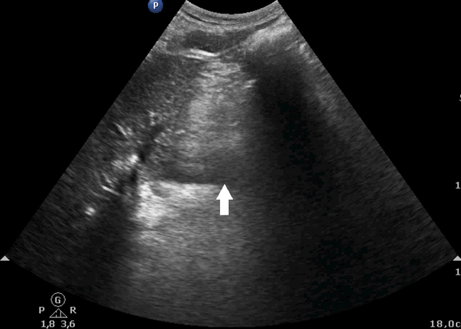
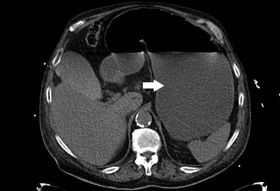
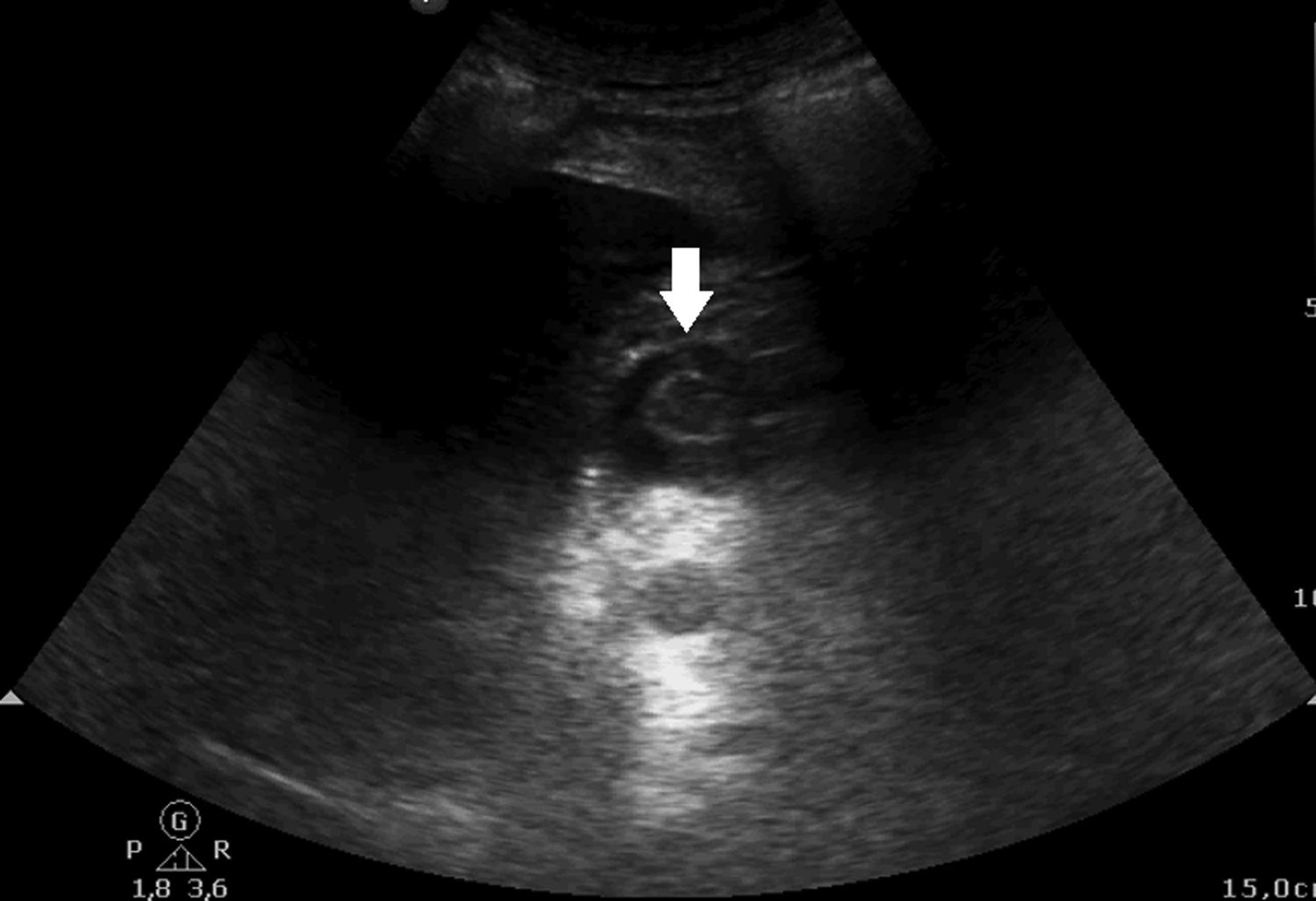
A few days later, the patient developed severe abdominal pain (visual analog score 8). The abdomen was seen to be bloated, with a tympanic percussion response. The nasogastric tube was positioned in aspiration, but produced no output. Abdominal ultrasound was performed, placing the probe transversely in the abdominal region between the epigastrium and the mesogastrium. The antrum was seen to be dilated, with content (echogenic) and no peristalsis (Fig. 1). The evaluation was completed with CAT, which confirmed the important gastric dilatation (Fig. 2) and the presence of the nasogastric tube in the esophagus. The tube was repositioned in aspiration, obtaining 1.5 liters of gastric content. The same abdominal ultrasound window subsequently confirmed an empty stomach through the target sign (Fig. 3). The patient was discharged after 6 days.
Ultrasound has become a crucial tool for dealing with a range of situations in the critically ill. Spending a few seconds to check correct positioning of the nasogastric tube in the stomach using ultrasound can avoid the use of invasive and needless techniques in our patients.
FundingThe authors declare that they have received no funding in relation to the present study.
Thanks are due to the Department of Intensive Care Medicine of Clínica Rotger.
Please cite this article as: Edroso Jarne PE, Monge Sola L, Vicho Pereira R. Mala colocación de la sonda nasogástrica diagnosticada por ecografía en paciente postoperado de cirugía abdominal. Med Intensiva. 2020;44:321–322.

