

Edited by: Juan Antonio Llompart-Pou - Specialist in Intensive Care Medicine, Doctorate in Health Sciences, Universitat Illes Balears. Department of Intensive Care Medicine, Trauma and Neurocritical ICU, Hospital Universitari Son Espases. Palma, Spain
Last update: April 2026
More infoNeurological prognosis of patients recovered from a cardiac arrest remains a challenge for Intensive Medicine specialists. Given the complexity of this scenario and the ensuing ethical dilemmas, current guidelines from the major scientific societies recommend a multimodal prognostic model for patients recovered from cardiac arrest. This model combines several clinical parameters, neurophysiological studies, such as electroencephalogram and somatosensory evoked potentials, and neuroimaging studies such as computed tomography scan and magnetic resonance imaging, as well as biomarkers for brain injury. More recently, several biomarkers associated with brain injury, originating from different regions of the brain, have been identified as potential prognostication tools within a multimodal approach. Based on the preliminary evidence gathered around them, several novel and promising biomarkers have been put forward. This literature review aims to examine four of them: ubiquitin carboxy-terminal hydrolase L1, glial fibrillary acidic protein, neurofilament light and tau protein.
El pronóstico neurológico de los pacientes recuperados tras una parada cardiorrespiratoria presenta un reto para el especialista en Medicina Intensiva. Dada la complicada naturaleza de este escenario y los conflictos éticos que genera, las recomendaciones actuales de las principales sociedades científicas sobre la atención del paciente recuperado de una parada cardiorrespiratoria recomiendan el uso de un modelo pronóstico multimodal. Para ello se emplea una combinación de parámetros clínicos, estudios neurofisiológicos como el electroencefalograma y los potenciales evocados somatosensoriales y estudios de neuroimagen como la tomografía computarizada y la resonancia magnética nuclear, así como biomarcadores de lesión cerebral. En los últimos años, varios biomarcadores de lesión cerebral originados en partes diferentes del cerebro han sido identificados como potenciales herramientas de valor pronóstico, dentro de una aproximación multimodal. Se han propuesto varios biomarcadores novedosos y prometedores alrededor de los cuales está surgiendo una evidencia preliminar. En esta revisión narrativa se pretende examinar cuatro de ellos: la ubiquitina carboxiterminal hidrolasa L1, la proteína ácida fibrilar de la glía, los neurofilamentos ligeros, y la proteína tau.
In patients who have recovered from cardiac arrest (CA), the hypoxic-ischemic brain injury resulting from the temporary circulatory arrest can lead to severe neurological complications. The clinical spectrum of this syndrome ranges from minimal or no functional sequelae to brain death due to cerebral edema. In many cases, we find an intermediate scenario in which the brain damage is not severe enough to cause death based on neurological criteria, but can lead to catastrophic functional consequences, such as a state of minimal consciousness or a persistent vegetative state. The identification of these cases presents a challenge to specialists in intensive care medicine, from both clinical and ethical perspectives. We know that some cases may have a favorable neurological outcome even weeks after the event. It is essential to recognize these cases to avoid making clinical decisions based on an inaccurate prognosis.
Recently published studies provide increasing evidence for incorporating new biomarkers into a multimodal strategy for establishing the neurological prognosis in this complex clinical setting. Blood biomarkers are readily available and are not affected by sedatives or neuromuscular blocking agents. Thanks to the latest analytical technologies, determining biomarkers in peripheral blood is not a risky technique. Samples collected for other routine purposes can also be used and compiled serially. Recent publications conclude that further studies are needed to continue evaluating the potential role of biomarkers in the clinical management of these patients.
The most recent guidelines from the European Resuscitation Council and the European Society of Intensive Care Medicine (ERC/ESICM) recommend using neurospecific enolase (NSE) as a prognostic tool 48−72 hours after the event.1 New biomarkers, including ubiquitin carboxy-terminal hydrolase L1 (UCH-L1), glial fibrillary acidic protein (GFAP), neurofilament light chain (NfL), and tau protein, are being studied as prognostic biomarkers. The emerging evidence is promising.2–4,17
This narrative review aims to concisely examine the latest published studies evaluating the prognostic value of the new biomarkers and their potential as prognostic tools in complex clinical settings.
MethodologyAn in-depth literature review was conducted using the PubMed, EMBASE, MEDLINE, and Scopus databases. Publications from 2014 to the present were reviewed, as more evidence on the study topic became available during that time. Descriptors included in the "Keywords" section were used. Neurological prognosis was defined as "favorable" for cases scoring between 1 and 2 on the Cerebral Performance Categories (CPC) scale, between 0 and 2 on the modified Rankin Scale (mRS), or 5 on the Glasgow Outcome Scale (GOS). All other cases were defined as "unfavorable".
All studies referring to new biomarkers were included if they were found to be related to neurological prognosis and/or analyzed the diagnostic accuracy comparing different biomarkers. The identified articles were analyzed based on their title, type of study, objectives, and conclusions. Due to the nature of the search, all the analyzed publications were descriptive studies, including systematic reviews and meta-analyses of these studies. Studies that did not meet these criteria and those published before 2014 were excluded. Likewise, publications involving patients under 18 years of age and those in which the functional scales used were not specified were excluded. After excluding the publications that did not meet the established criteria, a total of 18 studies were analyzed to prepare the sections of this narrative review.
ResultsUbiquitin Carboxy-Terminal Hydrolase L1(UCH-L1)The C-terminal fragment of UCH-L1 is a 26 kDa neuronal protein that is expressed mainly in neurons and neuroendocrine cells. UCH-L1 plays a crucial role in neuroaxonal stability and repair processes following brain injury. Specifically, it is involved in the ubiquitination process of proteins destined for degradation via the proteosome pathway.5 Therefore, UCH-L1 plays an important role in eliminating oxidized or misfolded proteins under both normal and pathological conditions. This marker is most commonly evaluated in traumatic brain injury (TBI), where its use has been approved by the United States FDA as an indicator of a favorable patient prognosis, helping to avoid unnecessary CT scans after non-severe head injury.6
Two studies have evaluated the use of UCH-L1 in patients who have recovered from cardiac arrest (CA), involving the same cohort of patients as in the TTM study.7,8 UCH-L1 was shown to accurately predict neurological outcomes after CA (area under the ROC between 0.85 and 0.87), performing significantly better than NSE at 24 and 48 h.
The prognostic estimation accuracy improved further with the addition of glial fibrillary acidic protein (GFAP) (area under the ROC between 0.90 and 0.91). At 72 h, both UCH-L1 and NSE behaved similarly, which can be explained by UCH-L1’s reported short half-life (< 12 h) compared to NSE. Further independent studies are required to define normal reference ranges and determine prognostic value alongside other neurological prognostic biomarkers after CA. Additionally, the non-specific expression of UCH-L1 from the pancreas and kidneys may pose a confounding factor for its use after CA.9
From these related studies, it is concluded that UCH-L1 specificity decreases over time, and especially after 48 h, when its accuracy is lower than that of NSE. However, the high specificity of UCH-L1suggests that it may play a role in fine-tuning the neurological prognosis following CA, especially within the first 24−48 hours after the event.9
Glial fibrillary acidic protein (GFAP)GFAP is a structural component of the intermediate filaments in the astrocyte cytoskeleton and is considered to be a specific marker of brain damage. GFAP synthesis is over-regulated after ischemic injury. It is believed that this protein is part of neuroprotective mechanisms and can lead to glial scarring phenomena.10 Determining GFAP levels in the blood can predict neurological outcomes after traumatic brain injury (TBI). Elevated levels have also been observed after CA, intracerebral hemorrhage, and ischemic stroke.10,11
In the context of post-CA encephalopathy, GFAP has only been investigated in three studies.8,11,12 GFAP appears to be more accurate in assessing the neurological prognosis at 48 and 72 h after the event than at earlier timepoints, with ROC curve values between 0.65 and 0.89. The largest published study, which was conducted in the TTM8 trial cohort, reported a threshold of 2,952 pg/mL with a 0% false-positive rate for predicting a poor neurological prognosis at 48 h post-CA; however, the sensitivity was generally low. The cut-offs considered in the remaining two studies were much lower (300 and 80 pg/mL), but the studies had important methodological differences compared to the TTM cohort study. In summary, the conclusions of the few published studies are not comparable due to methodological differences in data analysis and variability in results. In the Helwig et al.12 study, both NSE and GFAP showed modest accuracy results at 48 h, with NSE demonstrating higher sensitivity. However, in the Ebner et al.8 study, GFAP obtained showed specificity in predicting an adverse neurological outcome at each measurement timepoint (24, 48, and 72 h), at the cost of lower sensitivity. The ROC curve was clearly superior when GFAP and UCH-L1 results were analyzed together. Arctadeius et al.17 also observed in their study that elevated GFAP concentrations at 12 h post-CA in patients with a poor neurological prognosis had a predictive power similar to that of NSE at 48 h. This suggests an advantage to using GFAP as an early marker compared to NSE.17 Adding NFL to GFAP improved predictive accuracy at 12 and 48 h after out-of-hospital CA, but not at admission or after in-hospital CA. Cut-off points evaluated for poor neurological prognosis were 1,626 pg/mL at 12 h with a false-positive rate of less than 2% and a sensitivity of 34%.
According to the data analyzed in the studies evaluated in the present review, GFAP can be used to identify patients at an early stage with a higher probability of adverse neurological outcomes (12 and 24 h), based on its specificity values compared to NSE. Furthermore, the addition of NFL determination improves the overall accuracy at 12 and 48 h in out-of-hospital CA.
Tau proteinTau protein is a molecule that stabilizes microtubules in neuroaxonal processes. It is mainly found in the white matter of the central nervous system (CNS). Ischemic processes cause phosphorylation of the tau protein, resulting in its separation from microtubules. Hyperphosphorylated tau protein then aggregates into insoluble clumps, disrupting the transmission of electrochemical signals along the axon.13 The serum concentration of tau protein has been shown to be elevated after ischemic stroke and CA.14,15 Determination of tau protein concentration requires highly sensitive immunoassay analysis, which is available only in specialized laboratories.
To date, three studies have investigated the predictive potential of tau protein. The study by Mattsson et al., based on the TTM cohort, demonstrated predictive efficacy for poor neurological outcome after CA, especially in late determinations (48 and 72 h), with an area under the ROC curve of 0.90 and 0.91, respectively, and an accuracy comparatively higher than that of NSE.16 Two pilot studies reported a bimodal release of tau protein, with the late peak being significantly lower in patients with a good neurological outcome. This is explained by its relatively short elimination half-life of about 10 h, with late elevated levels reflecting persistent neuronal damage (second hit).4,14,17 The results of the study by Arctadeius et al.17 showed significantly elevated levels in patients with a poor prognosis from 12 h in out-of-hospital CA and at all timepoints in in-hospital CA. In all patients, regardless of neurological outcome, tau protein concentrations followed a bimodal pattern; in patients with a poor neurological outcome, late re-elevation of the marker was observed after 48 h. The predictive ability was highest at 48 h after out-of-hospital CA, with an area under the ROC curve of 0.93. At 48 h, levels above 35.5 pg/mL showed a false positive rate < 2% with a sensitivity of 86%.17 Compared to NSE, tau protein showed similar predictive values at 48 h.
Considering these results, it is clear that the usefulness of tau protein determination lies in the identification of patients with a poor neurological prognosis on a late basis at 72 h, with greater accuracy than NSE. In addition, due to the bimodal kinetics of tau protein release, those patients who show a second late increase at 48 h have a poorer neurological outcome, thus contributing to the identification of patients with a poor prognosis. As an additional result,17 the predictive value of tau protein at 12 h was found to be greater when GFAP measurement was added.
Neurofilament light chainNeurofilament chains (light, medium, heavy, and beta-internexin) are structural proteins that are uniquely expressed in neurons, predominantly in the myelinated axons of the white matter. Their exact function is currently unknown, although they are postulated to be an essential component of axonal development and nerve impulse conduction. Pathological processes result in the release of this structural component into the extracellular fluid, cerebrospinal fluid (CSF), and peripheral blood. Neurofilament light chains can also be detected in the fluids of healthy individuals and are released in a regular and age-dependent manner, doubling their normal concentration in 70-year-olds.18
In order to standardize measurements across countries, studies and centers, there is consensus that the determination of NFL levels using ultrasensitive and automated immunoassay techniques provides highly accurate quantification. As a result, cut-off points can be transferred and interpreted in a uniform manner across studies and centers. NFL has several key advantages as a biomarker, because it is not subject to major confounding factors and is not affected by pre-analytical variables or hemolysis.
In the context of hypoxic-ischemic brain injury, a very significant increase in NFL concentration has been observed, suggesting a possible prognostic role in this clinical context. This possibility was investigated in two recently published studies. The COMACARE study found that the median plasma NFL was >2,300 pg/mL in patients recovering from CA with a poor neurological prognosis, whereas the concentration was <20 pg/mL in patients with a good neurological outcome.3 Thus, NFL predicted the neurological prognosis in out-of-hospital CA with an area under the ROC curve of 0.98 at 24 h after the event. These results are consistent with those published by Moseby-Knappe et al., based on the TTM study cohort, who reported superior predictive accuracy with respect to other biomarkers such as tau protein and NSE in the 24-h window.4 In the study by Ryan and Hoiland, NFL concentration at 48 h showed an area under the ROC curve greater than that of the other markers in predicting neurological prognosis. On the other hand, an elevation of NFL above the cut-off points was observed in up to one-third of patients with a good neurological outcome, showing rather modest specificity.19 A more recent study, published by Arctaedius et al., found that NFL was superior to tau protein at 12 h in predicting poor prognosis, and the addition of NFL determination to tau protein levels improved predictive accuracy at all timepoints.17
In conclusion, these data suggest that early interpretation of normal NFL values (12−24 hours) may exclude a poor neurological prognosis and should be considered in the management of patients awaiting neurological recovery rather than in the identification of individuals with a poor prognosis.9
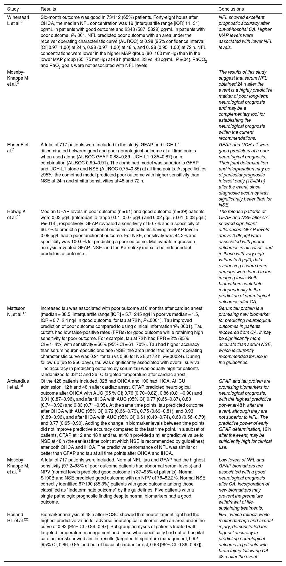
Table 1 summarizes the conclusions of the most relevant studies that analyzed the different biomarkers, as well as the cut-off points and other statistical data derived from the main studies that have been evaluated for this study.
Main studies analyzed, results, and conclusions.
| Study | Results | Conclusions |
|---|---|---|
| Wihersaari L et al.2 | Six-month outcome was good in 73/112 (65%) patients. Forty-eight hours after OHCA, the median NFL concentration was 19 (interquartile range [IQR] 11−31) pg/mL in patients with good outcome and 2343 (587−5829) pg/mL in patients with poor outcome, P<.001. NFL predicted poor outcome with an area under the receiver operating characteristic curve (AUROC) of 0.98 (95% confidence interval [CI] 0.97−1.00) at 24 h, 0.98 (0.97−1.00) at 48 h, and 0. 98 (0.95−1.00) at 72 h. NFL concentrations were lower in the higher MAP group (80−100 mmHg) than in the lower MAP group (65−75 mmHg) at 48 h (median, 23 vs. 43 pg/mL, P =.04). PaCO2 and PaO2 goals were not associated with NFL levels. | NFL showed excellent prognostic accuracy after out-of-hospital CA. Higher MAP levels were associated with lower NFL levels. |
| Moseby-Knappe M et al.3 | The results of this study suggest that serum NFL obtained 24 h after the event is a highly predictive marker of poor long-term neurological prognosis and may be a complementary tool for establishing the neurological prognosis within the current recommendations. | |
| Ebner F et al.7 | A total of 717 patients were included in the study. GFAP and UCH-L1 discriminated between good and poor neurological outcome at all time points when used alone (AUROC GFAP 0.88−0.89; UCH-L1 0.85−0.87) or in combination (AUROC 0.90−0.91). The combined model was superior to GFAP and UCH-L1 alone and NSE (AUROC 0.75−0.85) at all time points. At specificities ≥95%, the combined model predicted poor outcome with higher sensitivity than NSE at 24 h and similar sensitivities at 48 and 72 h. | GFAP and UCH-L1 were good predictors of a poor neurological prognosis. Their joint determination and interpretation may be of particular prognostic interest early (12−24 h) after the event, since diagnostic accuracy was significantly better than for NSE. |
| Helwig K et al.11 | Median GFAP levels in poor outcome (n = 61) and good outcome (n = 39) patients were 0.03 μg/L (interquartile range 0.01−0.07 μg/L) and 0.02 μg/L (0.01−0.03 μg/L; P=.014), respectively. GFAP revealed a sensitivity of 60.7% and a specificity of 66.7% to predict a poor functional outcome. All patients having a GFAP level > 0.08 μg/L had a poor functional outcome. For NSE, sensitivity was 44.3% and specificity was 100.0% for predicting a poor outcome. Multivariate regression analysis revealed GFAP, NSE, and the Karnofsky index to be independent predictors of outcome. | The release patterns of GFAP and NSE after CA showed significant differences. GFAP levels above 0.08 μg/l were associated with poorer outcomes in all cases, and in those with very high values (> 3 μg/l), data evidencing severe brain damage were found in the imaging tests. Both biomarkers contribute independently to the prediction of neurological outcomes after CA. |
| Mattsson N, et al.15 | Increased tau was associated with poor outcome at 6 months after cardiac arrest (median = 38.5, interquartile range [IQR] = 5.7−245 ng/l in poor vs median = 1.5, IQR = 0.7−2.4 ng/l in good outcome, for tau at 72 h, P<.0001). Tau improved prediction of poor outcome compared to using clinical information(P<.0001). Tau cutoffs had low false-positive rates (FPRs) for good outcome while retaining high sensitivity for poor outcome. For example, tau at 72 h had FPR = 2% (95% CI = 1−4%) with sensitivity = 66% (95% CI = 61−70%). Tau had higher accuracy than serum neuron-specific enolase (NSE; the area under the receiver operating characteristic curve was 0.91 for tau vs 0.86 for NSE at 72 h, P=.00024). During follow-up (up to 956 days), tau was significantly associated with overall survival. The accuracy in predicting outcome by serum tau was equally high for patients randomized to 33°C and 36°C targeted temperature after cardiac arrest. | Serum tau protein is a promising new biomarker for predicting neurological outcomes in patients recovered from CA. It may be significantly more accurate than serum NSE, which is currently recommended for use in the guidelines. |
| Arctaedius I et al.16 | Of the 428 patients included, 328 had OHCA and 100 had IHCA. At ICU admission, 12 h and 48 h after cardiac arrest, GFAP predicted neurological outcome after OHCA with AUC (95 % CI) 0.76 (0.70−0.82), 0.86 (0.81−0.90) and 0.91 (0.87−0.96), and after IHCA with AUC (95% CI) 0.77 (0.66−0.87), 0.83 (0.74−0.92) and 0.83 (0.71−0.95). At the same time points, tau predicted outcome after OHCA with AUC (95% CI) 0.72 (0.66−0.79), 0.75 (0.69−0.81), and 0.93 (0.89−0.96), and after IHCA with AUC (95% CI) 0.61 (0.49−0.74), 0.68 (0.56−0.79), and 0.77 (0.65−0.90). Adding the change in biomarker levels between time points did not improve predictive accuracy compared to the last time point. In a subset of patients, GFAP at 12 and 48 h and tau at 48 h provided similar predictive value to NSE at 48 h (the earliest time point at which NSE is recommended by guidelines) after both OHCA and IHCA. The predictive performance of NFL was similar or better than GFAP and tau at all time points after OHCA and IHCA. | GFAP and tau protein are promising biomarkers for neurological prognosis, with the highest predictive power at 48 h after the event, although they are not superior to NFL. The predictive power of early GFAP determination, 12 h after the event, may be sufficiently high for clinical use. |
| Moseby-Knappe M, et al.18 | A total of 717 patients were included. Normal NFL, tau and GFAP had the highest sensitivity (97.2−98% of poor outcome patients had abnormal serum levels) and NPV (normal levels predicted good outcome in 87−95% of patients). Normal S100B and NSE predicted good outcome with an NPV of 76−82.2%. Normal NSE correctly identified 67/190 (35.3%) patients with good outcome among those classified as "indeterminate outcome" by the guidelines. Five patients with a single pathologic prognostic finding despite normal biomarkers had a good outcome. | Low levels of NFL and GFAP biomarkers are associated with a good neurological prognosis after CA. Incorporation of new biomarkers may prevent the premature withdrawal of life-sustaining treatments. |
| Hoiland RL et al.22 | Biomarker analysis at 48 h after ROSC showed that neurofilament light had the highest predictive value for adverse neurological outcome, with an area under the curve of 0.92 (95% CI, 0.84−0.97). Subgroup analyses of patients treated with targeted temperature management and those who specifically had out-of-hospital cardiac arrest showed similar results (targeted temperature management, 0.92 [95% CI, 0.86−0.95] and out-of-hospital cardiac arrest, 0.93 [95% CI, 0.86−0.97]). | NFL, which reflects white matter damage and axonal injury, demonstrated the highest accuracy in predicting neurological outcome in patients with brain injury following CA 48 h after the event. |
The management of CA and its sequelae represents a significant use of healthcare resources and has a major impact on the quality of life of patients and their families. While there have been advances in immediate care and in the chain of survival (activation, quality cardiopulmonary resuscitation [CPR], early defibrillation, advanced life support, etc.), the development of therapeutic interventions aimed at post-return of spontaneous circulation brain injury has experienced little progress. Guidelines and consensus documents provide few new recommendations for the treatment of brain injury, most of which have a low level of evidence. The lack of progress in this area is an obstacle for the International Liaison Committee on Resuscitation (ILCOR).20,21 From a future research perspective, the Core Outcome Set for Cardiac Arrest (COSCA21 initiative has identified a framework in which outcome indicators such as functional status at discharge and quality of life should become more relevant.22
In light of recent studies, it seems clear that the new neurospecific biomarkers may be helpful in the complex clinical context of brain injury after CA. On the one hand, the collection and processing of samples does not pose a risk, since they can be obtained from peripheral blood, and on the other hand, the analytical technologies allow standardization of cut-off points and transferability of conclusions from one study to another. The particular kinetics of each marker may facilitate the use of one (or a combination of) biomarkers in each clinical setting, depending on whether sensitivity or specificity is prioritized at each timepoint. For example, normal NFL levels in the early stages (12–24 h) may be relatively certain to exclude a poor neurological prognosis and thus identify patients who are likely to awake later, or may be a favorable factor in the decision to prolong life support.
Similarly, and in line with the conclusions of recently published studies, it is true that the current evidence is preliminary and mainly derived from studies that are heterogeneous in terms of methodology and definitions. The cut-off values used to define outcomes show wide variability, and the analytical technology developed over the last decade may imply substantial differences between studies. Despite these limitations, there is a growing interest in new biomarkers as part of a multimodal diagnostic and prognostic approach that focuses on the functional status and quality of life of patients with TBI after return of spontaneous circulation in the medium to long term.
Financial supportThis work has received no funding.
Declaration of Generative AI and AI-assisted technologies in the writing processNo AI has been used for this review.
Author contributions CRediT authorship contribution statementJavier Puerma Jiménez: Acquisition, analysis, and interpretation of data and drafting of the article.
Ana Carrasco Cáliz: Critical review of the intellectual content.
José Miguel Pérez Villares: Final approval of the submitted version.
Antonio Cárdenas Cruz: Conception and design of the study.
The authors declare that they have no conflicts of interest in relation to this review.