


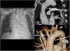
This is the case of a 50-year-old man without a significant past medical history treated outpatiently after sustaining an aggression with a bladed weapon at sternal notch level and who required prehospital intubation due to agitation. Afterwards, he is treated at the ICU where hemodynamic impairment is confirmed followed by the need to activate the massive bleeding protocol. The thoracic x-ray performed reveals the presence of mediastinal widening suggestive of aortic lesion (Fig. 1.1). The e-FAST protocol ultrasound revealed the presence of left side hemothorax with placement of thoracic drainage. After stabilizing the patient, he underwent a CT scan that confirmed the presence of 1 transfixed wound at aortic arch level with presence of pseudoaneurysms. Entry site: superior vena cava. Exit site: through the posterior-medial region (marked with white arrows in Fig. 1.2 and 1.3). These findings led to an emergency procedure that included the suture of the aortic upper wound. The patient’s progression at the ICU and at the hospital conventional ward was satisfactory.
Funding
None whatsoever.
We wish to thank the radiology unit at Hospital Universitario 12 de Octubre for their collaboration.

