


This is the case of an 80-year-old man with no relevant past medical history regarding the case he is a admitted for to the ICU after being run over by a vehicle. The patient had a Glasgow Coma Score (GCS) of 7 points, respiratory failure with flail chest, and showed hemodynamic instability. Upon arrival at the emergency department, the whole-body CT scan performed revealed multiple fractures of bilateral rib arcs, but no images consistent with esophagogastric pneumatosis.
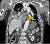
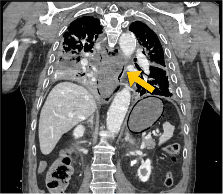
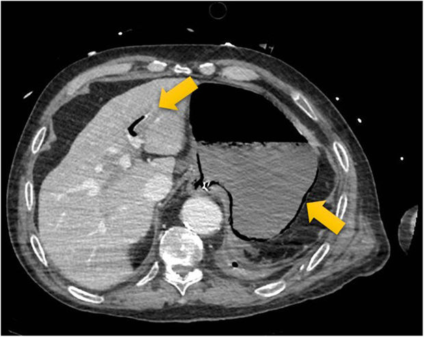
The patient was admitted to the ICU for continued care but had progressive worsening with refractory hypoxemia. A new imaging modality was performed 24h later that showed significant gastric chamber distention with pneumatosis in its wall and in the esophageal wall (Figs. 1 and 2) associated with portal pneumatosis in the main and left branches (Fig. 3). Given the patient's clinical context, the findings, although nonspecific, were indicative of the presence of gastric emphysema.
Conflict of interest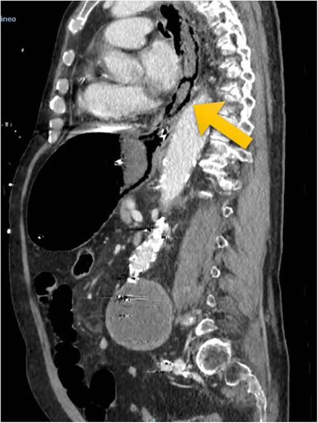
None whatsoever.
FundingNone reported.

