


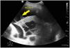
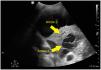
A 52-year-old patient with a history of acquired immunodeficiency syndrome (AIDS) was admitted to our Unit due to Streptococcus pneumoniae community-acquired pneumonia. The chest radiographs evidenced right basal infiltration, and the computed tomography (CT) study showed necrotizing pneumonia with a probable incipient lung abscess. The clinical course was poor, with hypoxemia refractory to cycles in prone decubitus. Pulmonary ultrasound revealed pleural effusion containing hyperechogenic fibrous tracts (plankton sign) (Fig. 1, arrow 1), right basal infiltration with hyperechogenic parenchyma secondary to hepatization, air bronchogram (Fig. 2, arrow 2), and well delimited internal hypoechogenic trabeculate zones consistent with necrotizing pneumonia and lung abscess (Fig. 2, arrow 3).
Conflicts of interest

The authors, L. Javier Pérez-Bazaga, Carlos Ávila-Sansegundo and María Ángeles Santiago-Triviño declare that they have no conflicts of interest.
FundingThe authors have received no funding for the present study.

