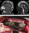
A 72-year-old female patient with history of high blood pressure presented to the emergency with right hemiparesis, aphasia and seizures. Brain computed tomography did not find hemorrhagic lesions and brain magnetic resonance imaging showed no vascular lesions but revealed laminar subdural collection in the right parietal convexity with restriction on the diffusion sequence together with multiple areas of restriction in the subarachnoid space on the bihemispheric convexity (Fig. 1, Panel A and B). It evolved with Glasgow coma scale 8/15, fever and saturation 87% due to aspiration, proceeding to endotracheal intubation. Blood cultures and lumbar puncture were performed. It showed glucose 49mg/dl (serum glucose 484mg/dl), protein level of 946g/dl, leukocytes 3744/mm3 (95% neutrophils), 1.000erythrocytes/mm3. Blood and cerebrospinal fluid cultures revealed the presence of Streptococcus pneumoniae. Neurosurgical intervention was decided with craniectomy and drainage of meningeal empyema (Panel C). The patient completed 8 weeks of ceftriaxone with good clinical outcome.
array:23 [ "pii" => "S0210569119303031" "issn" => "02105691" "doi" => "10.1016/j.medin.2019.11.011" "estado" => "S300" "fechaPublicacion" => "2022-03-01" "aid" => "1434" "copyright" => "Elsevier España, S.L.U. y SEMICYUC" "copyrightAnyo" => "2019" "documento" => "article" "crossmark" => 1 "subdocumento" => "sco" "cita" => "Med Intensiva. 2022;46:176" "abierto" => array:3 [ "ES" => true "ES2" => true "LATM" => true ] "gratuito" => true "lecturas" => array:1 [ "total" => 0 ] "itemSiguiente" => array:18 [ "pii" => "S0210569119303043" "issn" => "02105691" "doi" => "10.1016/j.medin.2019.12.006" "estado" => "S300" "fechaPublicacion" => "2022-03-01" "aid" => "1435" "copyright" => "Elsevier España, S.L.U. y SEMICYUC" "documento" => "article" "crossmark" => 1 "subdocumento" => "sco" "cita" => "Med Intensiva. 2022;46:177-8" "abierto" => array:3 [ "ES" => true "ES2" => true "LATM" => true ] "gratuito" => true "lecturas" => array:2 [ "total" => 2 "HTML" => 2 ] "es" => array:11 [ "idiomaDefecto" => true "cabecera" => "<span class="elsevierStyleTextfn">Imágenes en Medicina Intensiva</span>" "titulo" => "Disección coronaria espontánea, e idiopática, del tronco coronario izquierdo en mujer joven y sana" "tienePdf" => "es" "tieneTextoCompleto" => "es" "paginas" => array:1 [ 0 => array:2 [ "paginaInicial" => "177" "paginaFinal" => "178" ] ] "titulosAlternativos" => array:1 [ "en" => array:1 [ "titulo" => "Spontaneous and idiopathic coronary dissection of the left main coronary artery occurring in a young and healthy woman" ] ] "contieneTextoCompleto" => array:1 [ "es" => true ] "contienePdf" => array:1 [ "es" => true ] "resumenGrafico" => array:2 [ "original" => 0 "multimedia" => array:6 [ "identificador" => "fig0005" "etiqueta" => "Figura 1" "tipo" => "MULTIMEDIAFIGURA" "mostrarFloat" => true "mostrarDisplay" => false "figura" => array:1 [ 0 => array:4 [ "imagen" => "gr1.jpeg" "Alto" => 947 "Ancho" => 2508 "Tamanyo" => 316386 ] ] ] ] "autores" => array:1 [ 0 => array:2 [ "autoresLista" => "D.I. Gentille Lorente, M. Faiges Borrás, C. Moreno Ambroj" "autores" => array:3 [ 0 => array:2 [ "nombre" => "D.I." "apellidos" => "Gentille Lorente" ] 1 => array:2 [ "nombre" => "M." "apellidos" => "Faiges Borrás" ] 2 => array:2 [ "nombre" => "C." "apellidos" => "Moreno Ambroj" ] ] ] ] ] "idiomaDefecto" => "es" "EPUB" => "https://multimedia.elsevier.es/PublicationsMultimediaV1/item/epub/S0210569119303043?idApp=WMIE" "url" => "/02105691/0000004600000003/v1_202202250826/S0210569119303043/v1_202202250826/es/main.assets" ] "itemAnterior" => array:18 [ "pii" => "S0210569121001121" "issn" => "02105691" "doi" => "10.1016/j.medin.2021.05.007" "estado" => "S300" "fechaPublicacion" => "2022-03-01" "aid" => "1674" "copyright" => "Elsevier España, S.L.U. y SEMICYUC" "documento" => "simple-article" "crossmark" => 1 "subdocumento" => "cor" "cita" => "Med Intensiva. 2022;46:175" "abierto" => array:3 [ "ES" => true "ES2" => true "LATM" => true ] "gratuito" => true "lecturas" => array:1 [ "total" => 0 ] "en" => array:10 [ "idiomaDefecto" => true "cabecera" => "<span class="elsevierStyleTextfn">Letter to the Editor</span>" "titulo" => "Reply to “Decibans: it is time to weight the evidence about diagnostic accuracy”" "tienePdf" => "en" "tieneTextoCompleto" => "en" "paginas" => array:1 [ 0 => array:1 [ "paginaInicial" => "175" ] ] "titulosAlternativos" => array:1 [ "es" => array:1 [ "titulo" => "Respuesta a «Decibanes: es hora de pesar la evidencia sobre exactitud diagnóstica»" ] ] "contieneTextoCompleto" => array:1 [ "en" => true ] "contienePdf" => array:1 [ "en" => true ] "autores" => array:1 [ 0 => array:2 [ "autoresLista" => "C. Ramos-Vera" "autores" => array:1 [ 0 => array:2 [ "nombre" => "C." "apellidos" => "Ramos-Vera" ] ] ] ] ] "idiomaDefecto" => "en" "EPUB" => "https://multimedia.elsevier.es/PublicationsMultimediaV1/item/epub/S0210569121001121?idApp=WMIE" "url" => "/02105691/0000004600000003/v1_202202250826/S0210569121001121/v1_202202250826/en/main.assets" ] "en" => array:12 [ "idiomaDefecto" => true "cabecera" => "<span class="elsevierStyleTextfn">Images in Intensive Medicine</span>" "titulo" => "Meningeal empyema" "tieneTextoCompleto" => true "paginas" => array:1 [ 0 => array:1 [ "paginaInicial" => "176" ] ] "autores" => array:1 [ 0 => array:4 [ "autoresLista" => "N.A. Gemelli, N.M. Ciarrocchi, E. San Roman" "autores" => array:3 [ 0 => array:4 [ "nombre" => "N.A." "apellidos" => "Gemelli" "email" => array:1 [ 0 => "nicolas.gemelli@hospitalitaliano.org.ar" ] "referencia" => array:1 [ 0 => array:2 [ "etiqueta" => "<span class="elsevierStyleSup">*</span>" "identificador" => "cor0005" ] ] ] 1 => array:2 [ "nombre" => "N.M." "apellidos" => "Ciarrocchi" ] 2 => array:2 [ "nombre" => "E." "apellidos" => "San Roman" ] ] "afiliaciones" => array:1 [ 0 => array:2 [ "entidad" => "Adult Intensive Care Department, Hospital Italiano de Buenos Aires, Argentina" "identificador" => "aff0005" ] ] "correspondencia" => array:1 [ 0 => array:3 [ "identificador" => "cor0005" "etiqueta" => "⁎" "correspondencia" => "Corresponding author." ] ] ] ] "titulosAlternativos" => array:1 [ "es" => array:1 [ "titulo" => "Empiema meningeo" ] ] "resumenGrafico" => array:2 [ "original" => 0 "multimedia" => array:6 [ "identificador" => "fig0005" "etiqueta" => "Figure 1" "tipo" => "MULTIMEDIAFIGURA" "mostrarFloat" => true "mostrarDisplay" => false "figura" => array:1 [ 0 => array:4 [ "imagen" => "gr1.jpeg" "Alto" => 916 "Ancho" => 800 "Tamanyo" => 160384 ] ] ] ] "textoCompleto" => "<span class="elsevierStyleSections"><p id="par0005" class="elsevierStylePara elsevierViewall">A 72-year-old female patient with history of high blood pressure presented to the emergency with right hemiparesis, aphasia and seizures. Brain computed tomography did not find hemorrhagic lesions and brain magnetic resonance imaging showed no vascular lesions but revealed laminar subdural collection in the right parietal convexity with restriction on the diffusion sequence together with multiple areas of restriction in the subarachnoid space on the bihemispheric convexity (<a class="elsevierStyleCrossRef" href="#fig0005">Fig. 1</a>, Panel A and B). It evolved with Glasgow coma scale 8/15, fever and saturation 87% due to aspiration, proceeding to endotracheal intubation. Blood cultures and lumbar puncture were performed. It showed glucose 49<span class="elsevierStyleHsp" style=""></span>mg/dl (serum glucose 484<span class="elsevierStyleHsp" style=""></span>mg/dl), protein level of 946<span class="elsevierStyleHsp" style=""></span>g/dl, leukocytes 3744/mm<span class="elsevierStyleSup">3</span> (95% neutrophils), 1.000<span class="elsevierStyleHsp" style=""></span>erythrocytes/mm<span class="elsevierStyleSup">3</span>. Blood and cerebrospinal fluid cultures revealed the presence of <span class="elsevierStyleItalic">Streptococcus pneumoniae</span>. Neurosurgical intervention was decided with craniectomy and drainage of meningeal empyema (Panel C). The patient completed 8 weeks of ceftriaxone with good clinical outcome.</p><elsevierMultimedia ident="fig0005"></elsevierMultimedia></span>" "pdfFichero" => "main.pdf" "tienePdf" => true "multimedia" => array:1 [ 0 => array:6 [ "identificador" => "fig0005" "etiqueta" => "Figure 1" "tipo" => "MULTIMEDIAFIGURA" "mostrarFloat" => true "mostrarDisplay" => false "figura" => array:1 [ 0 => array:4 [ "imagen" => "gr1.jpeg" "Alto" => 916 "Ancho" => 800 "Tamanyo" => 160384 ] ] ] ] ] "idiomaDefecto" => "en" "url" => "/02105691/0000004600000003/v1_202202250826/S0210569119303031/v1_202202250826/en/main.assets" "Apartado" => array:4 [ "identificador" => "440" "tipo" => "SECCION" "es" => array:2 [ "titulo" => "Imágenes en medicina intensiva" "idiomaDefecto" => true ] "idiomaDefecto" => "es" ] "PDF" => "https://static.elsevier.es/multimedia/02105691/0000004600000003/v1_202202250826/S0210569119303031/v1_202202250826/en/main.pdf?idApp=WMIE&text.app=https://www.medintensiva.org/" "EPUB" => "https://multimedia.elsevier.es/PublicationsMultimediaV1/item/epub/S0210569119303031?idApp=WMIE" ]