Journal Information
Vol. 46. Issue 12.
Pages 731-732 (December 2022)
Share
Share
Download PDF
More article options
Vol. 46. Issue 12.
Pages 731-732 (December 2022)
Images in Intensive Medicine
Full text access
Echocardiography features of non-compaction cardiomyopathy
Imagen ecográfica de miocardiopatía no compactada (MNC)
Visits
806
R. Molina Lobo
Corresponding author
rosmolinalobo@msn.com

Corresponding author.
, I. Conejo Márquez, C. Hermosa Gelbard
Servicio de Medicina Interna, Hospital Universitario de Henares, Coslada, Madrid, Spain
This item has received
Article information
Full Text
Download PDF
Statistics
Figures (3)
Show moreShow less
Additional material (1)
Full Text

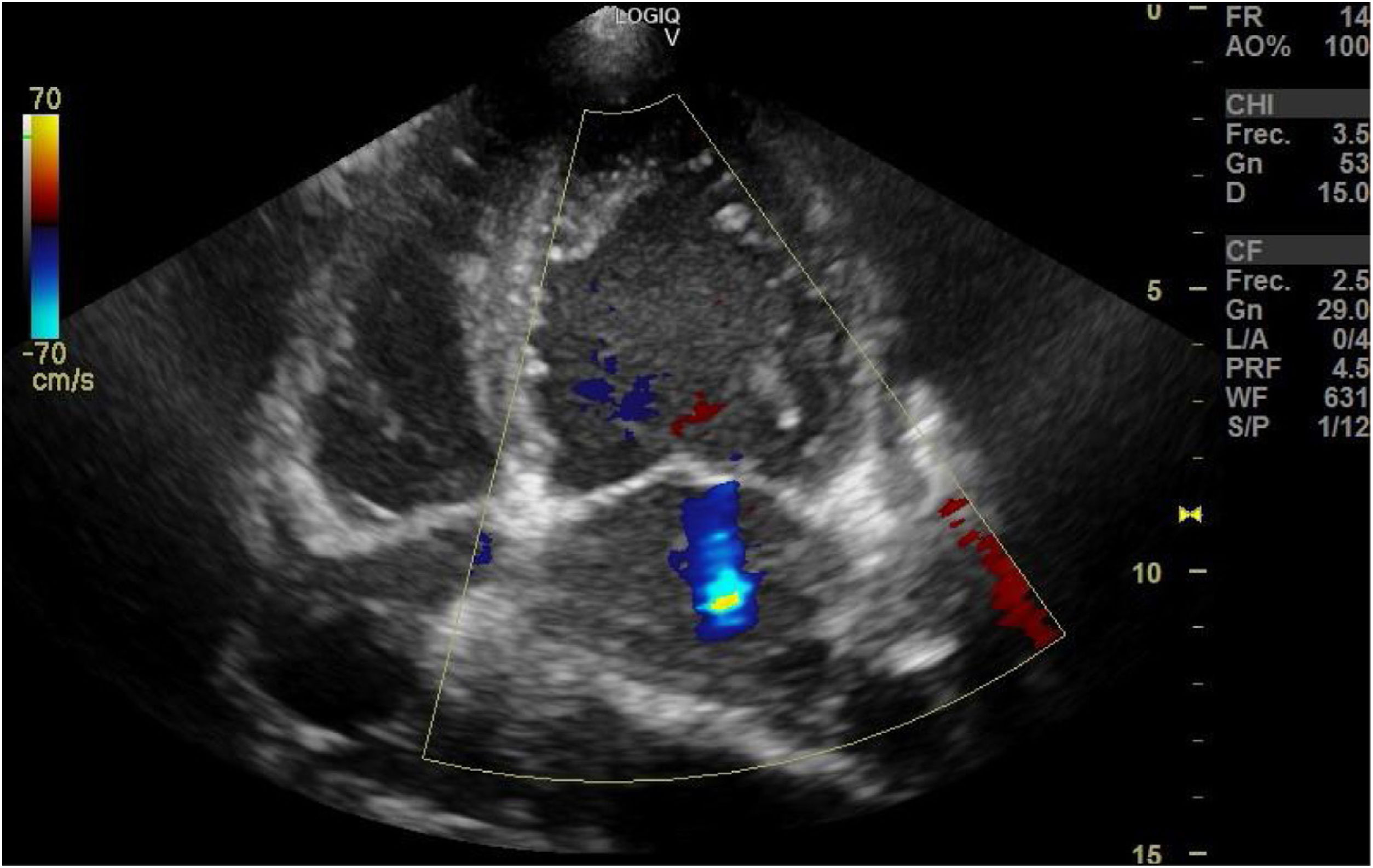
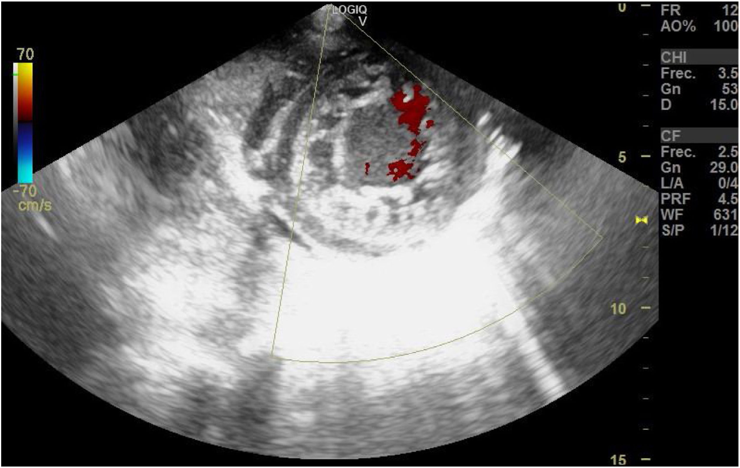
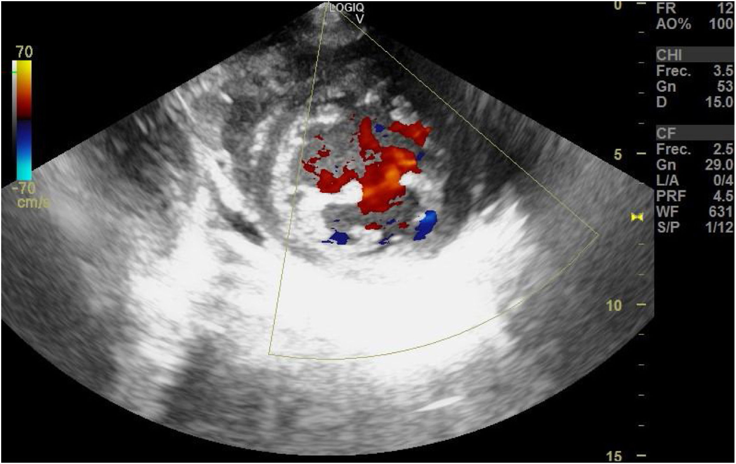
Non-compaction cardiomyopathy is a rare entity associated with complications such as heart failure, cardioembolic phenomena, arrhythmias, and sudden death. Etiology is not fully understood, but it has a genetic base. Its diagnosis is increasing due to the use of echocardiography and is characterized by the thickening of left ventricular wall with prominences and recesses with absence of compacted myocardium while 2 different layers can be distinguished: compacted myocardium and non-compaction myocardium (Fig. 1). On the color Doppler echocardiography blood flow can be seen flowing between such crypts without communication with the coronary system (Figs. 2 and 3) with the possibility of causing emboli.

Figure 1
(0.27MB).
Figure 2
(0.29MB).
Figure 3
(0.3MB).

We are providing echocardiographic images of a 62-year-old woman with clinical signs of respiratory failure due to ARF in the ventricular dysfunction setting (Video).

Appendix A
Supplementary data

The following is Supplementary data to this article:

(0.75MB)

Copyright © 2020. Elsevier España, S.L.U. and SEMICYUC
Idiomas
Medicina Intensiva (English Edition)
Article options
Tools
Supplemental materials
es en

¿Es usted profesional sanitario apto para prescribir o dispensar medicamentos?

Are you a health professional able to prescribe or dispense drugs?