


Thoracic surgery has undergone significant advances in recent years related to anesthetic and surgical techniques and the prevention and management of complications related to the procedure. This has allowed improvements in patient clinical outcomes in surgeries of this kind. Despite the above, thoracic surgery, especially related to pulmonary resection, is not without risk, and is associated to considerable morbidity and mortality. Fast track or enhanced recovery after anesthesia protocols, minimally invasive surgery, and intraoperative anesthetic management improve the prognosis and safety of thoracic surgery. Patients in the postoperative period of major thoracic surgery require intensive surveillance, especially the first 24–72h after surgery. Admission to the ICU is especially recommended in those patients with comorbidities, a reduced cardiopulmonary reserve, extensive lung resections, and those requiring support due to life-threatening organ failure. During the postoperative period, intensive cardiorespiratory monitoring, proper management of thoracic drainage, aggressive pain control (multimodal analgesia and regional anesthetic techniques), nausea and multimodal rehabilitation are key elements for avoiding adverse events. Medical complications include respiratory failure, arrhythmias, respiratory infections, atelectasis and thromboembolic lung disease. The most frequent surgical complications are hemothorax, chylothorax, bronchopleural fistula and prolonged air leakage. The multidisciplinary management of these patients throughout the perioperative period is essential in order to ensure the best surgical outcomes.
La cirugía torácica ha experimentado importantes avances en los últimos años relacionados con las técnicas anestésicas y quirúrgicas y la prevención y el manejo de las complicaciones relacionadas con el procedimiento. Esto ha permitido mejorar los resultados clínicos de los pacientes sometidos a este tipo de intervención. A pesar de ello, los procedimientos de cirugía torácica, especialmente los relacionados con la resección pulmonar, no están exentos de riesgo, con una morbimortalidad asociada considerable. Los protocolos Fast track o Enhanced recovery after anesthesia, la cirugía mínimamente invasiva y el manejo anestésico intraoperatorio mejoran el pronóstico y la seguridad de los procesos de cirugía torácica. Los pacientes postoperados de cirugía torácica mayor requieren una vigilancia intensiva, especialmente las primeras 24-72h del postoperatorio inmediato. El ingreso en la UCI se recomienda especialmente en los pacientes con comorbilidad, con reserva cardiopulmonar reducida, con resecciones pulmonares extensas y en los que requieren soporte por fallo de algún órgano con riesgo vital. Durante el periodo postoperatorio la monitorización intensiva cardiorrespiratoria, el manejo adecuado de los drenajes torácicos, el control agresivo del dolor (analgesia multimodal y técnicas anestésicas regionales), las náuseas y la rehabilitación multimodal son elementos claves para evitar eventos adversos. Entre las complicaciones médicas destacan la insuficiencia respiratoria, las arritmias, las infecciones respiratorias, las atelectasias y la enfermedad pulmonar tromboembólica. Las complicaciones quirúrgicas más frecuentes son el hemotórax, el quilotórax, la fístula broncopleural y la fuga aérea prolongada. El manejo multidisciplinar de estos pacientes durante todo el periodo perioperatorio es esencial para asegurar los mejores resultados quirúrgicos.
The current new anesthesia induction techniques and the advances made in perioperative care and surgical procedures facilitate the performance of major interventions with fewer intraoperative and postoperative complications.
Forty percent of in-hospital adverse events are associated with surgical procedures,1 which means that every year up to 7 million patients will suffer severe complications during or immediately after surgery, and 1 million will eventually die. According to studies conducted in developing countries, the mortality rate of major surgeries is between 5% and 10%.
The risk of perioperative adverse events depends on the patient's condition prior to the surgery, the prevalence of comorbidities, the urgency, the magnitude, type, and duration of the surgery.
Over the last few decades we have seen 2 major advances that have impacted surgical outcomes significantly: minimally invasive surgery and multimodal rehabilitation programs also known as the Fast track o Enhanced Recovery After Anesthesia (ERAS) protocol. The goal of both strategies is to reduce surgical aggressiveness, thus facilitating postoperative recovery.2
Thoracic surgeryThoracic surgery is a highly complex surgery performed using different procedures: mediastinoscopy, video-assisted thoracoscopy, sympathectomy, pulmonary wedge resection, segmentectomy, lobectomy, pneumectomy, volume reduction surgery, thoracic wall surgery, tracheal surgery, and esophageal surgery. Patients treated with lung-resection surgery are often patients with major comorbidities such as chronic obstructive pulmonary disease, heart disease, and in most cases, they are cancer patients.
Other factors associated with the intraoperative management of these patients like one-lung ventilation favor the intraoperative and postoperative pathophysiological disturbances, and the appearance of respiratory and cardiovascular complications that add up to the complications traditionally associated with surgery.
Procedural mortality associated with thoracic surgery especially in parenchymatous resections has dropped below 5% in recent series. The Society of Thoracic Surgeons General Thoracic Surgery Database has a registry of 19903 patients and reports a 1.8% mortality rate, 5-day hospital stays on average, and lung complication rates of 18.5%.3
Periprocedural managementIn surgery multimodal rehabilitation is the combination of evidence-based medicine-sustained perioperative strategies. Its main goal is to improve recovery after surgery. These strategies include the Fast track or ERAS protocols. Until now, the greater scientific evidence coming from the ERAS protocols derives from studies conducted with patients treated with oncological colorectal cancer surgery. The ERAS protocols reduced mortality, improve postoperative recovery, and reduced hospital stay.4
Multidisciplinary strategy combines training the patient before surgery, reducing post-traumatic stress with new anesthesia, analgesia, and drug induction techniques, minimally invasive surgery, aggressive postoperative rehabilitation, and reviewing the classic principles of postoperative care (transducer, drainage, catheter, etc.) to avoid complications, improve the patient's early recovery, and thus, reduce hospital stay.5
Although this multimodal approach has not been implemented extensively in thoracic surgery, there are initiatives that have shown good results.6 During the entire lung resection process, European studies recommend early intubation, mobilization, use of multimodal analgesia, and the early withdrawal of thoracic tubes. The European Society of Thoracic Surgeons has recently published its recommendations for the management of patients undergoing lung surgery (Table 1).7
Recommendations for the perioperative management of patients treated with lung surgery according to the European Society of Thoracic Surgeons.
| Preparation |
| Detailed information and recommendations to quit smoking and avoid alcohol consumption at least 4 weeks prior to the intervention |
| Nutritional supplements for malnourished patients |
| Anemia identification and correction |
| Preconditioning of patients with borderline pulmonary functional reserve |
| Preoperative |
| Preoperative fasting: |
| Water up to 2h prior to the induction of anesthesia and solid food up to 6h prior to the induction of anesthesia |
| Oral overload of carbon hydrates prior to the intervention |
| The routine use of sedatives to manage anxiety prior to the intervention is not recommended |
| Perioperative |
| Antibiotic prophylaxis and skin washing |
| Use of chlorhexidine solutions to prepare the skin |
| Continuous control of temperature and prevention of hypothermia |
| Lung protective mechanical ventilation especially in cases of one lung ventilation |
| Use of combined anesthesia techniques (regional and general) and half-life for anesthetic drugs |
| Combination of non-pharmacological and pharmacological measures for postoperative nausea and vomiting (PONV) prevention |
| The use of epidural anesthesia for the management of pain and avoidance of opioids is recommended |
| The analgesia provided by paravertebral block is equivalent to that provided by epidural anesthesia |
| The use of conventional analgesia with paracetamol and NSAIDs is recommended except for contraindications |
| Dexamethasone can be administered to reduce PONV |
| Avoid highly restrictive or liberal protocols of serum therapy |
| The use of balanced solutions is preferred over saline serum solutions at 0.9% |
| Oral tolerance and serum therapy withdrawal will be initiated as soon as possible |
| Prevention of atrial fibrillation: |
| Reintroduce beta-blockers as soon as possible in patients on chronic treatment |
| Assess the use of preoperative diltiazem or postoperative amiodaron in high-risk patients |
| External pleural drainage: |
| Do not apply external aspiration as a general rule |
| Assess the removal of drainage systems as soon as possible even in cases with 450mL/day drains |
| The use of a single drainage tube for is recommended |
| Avoid the use of an unnecessary vesical catheter and proceed with early withdrawal if used |
| Early mobilization within the first 24h |
| Thromboembolic prophylaxis in patients treated with major lung surgery. In very high-risk cases assess whether to keep on administering prophylaxis for up to 4 weeks |
Adapted from Batchelor et al.7.
The concept of «minimally invasive surgery» refers to established surgical procedures performed remotely in a confined space. Video-assisted thoracic surgery (CTAV or VATS) or video-assisted thoracoscopy is the most advanced minimally invasive surgery. The benefits of this approach are less trauma, fewer complications, faster recovery time, work insertion and social inclusion, and better cosmetic results.
There are very few data on VATS procedures because no large randomized studies have been conducted to compare this technique with lung resection through open surgery. A meta-analysis showed that VATS lobectomy was associated with a non-significant difference in mortality, but with significantly lower perioperative rates of morbidity, pneumonia, and atrial fibrillation.8
It has been proven that this is a safe and satisfactory procedure from the oncological standpoint even for locally advanced tumors such as lung tumor resection with thoracic wall involvement.9
Admission criteria in intensive care unitsAlthough the systematic admission in intensive care units (ICU) of patients treated with thoracic surgery has been questioned, especially in low-risk patients, those treated with major thoracic surgery can require admission for 24–72h in these units.
The clinical practice guidelines of the European Respiratory Society/European Society of Thoracic Surgeons10 and recent studies11 provide data on criteria that may be a guide on what the best destination of these patients may be depending on the risks, benefits, and resources available. ICU admission is especially recommended in patients with comorbidities, a low cardiopulmonary reserve, extensive lung resections, and those who require support due to organ failure with life-threatening risk for the patient (Table 2).12
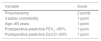
The use of predictive indices can also be useful when it comes to deciding where the patient should be transferred to after receiving thoracic surgery. One of these indices measures the probability of ICU admission depending on the complications experienced after major lung resection.13 It uses different variables and assigns different scores (Table 3). Patients with scores ≥4 would have a high risk of experiencing serious complications and, therefore, would be eligible for ICU admission.
Variables to predict the possibility of serious complications that may require urgent ICU admission.
| Variable | Score |
|---|---|
| Pneumectomy | 2 points |
| Cardiac comorbidity | 1 point |
| Age >65 years | 1 point |
| Postoperative predictive FEV1 <65% | 1 point |
| Postoperative predictive DLCO <50% | 1 point |
DLCO, diffusing capacity of lung for carbon monoxide; FEV1, maximal forced expiratory volume in the first second.
Intensive cardiorespiratory monitorization (electrocardiogram, blood pressure, heart rate, respiratory rate, oxygen saturation measured through pulse oximetry, and periodic controls of arterial-blood gas test) are basic elements in the management of these patients. In unstable patients or patients with complications, a more aggressive management may be necessary by increasing the level of monitorization. The use of fluids has been associated with the appearance of acute lung injury, which is why careful use is recommended in these patients during pre- and post-operative management with strict control of diuresis and renal function.14
The management of thoracic drainage is one characteristic procedures in these patients and recent studies with new pieces of equipment have questioned conventional management.15 The use of a single drainage can reduce the appearance of pain.16 Portable suction units instead of the traditional wall-suction systems allow mobilization and even early walking. The routine use of thoracic X-rays does not affect the management of thoracic tubes and it should be used selectively. There is no consensus on the indication for the removal of drainage systems based on the amount of fluid produced. In most cases they can be safely removed within the first 24h. Despite randomized studies and meta-analyses, the optimal negative pressure or its superiority have not been established regarding water-seal drainage. With the use of new digital pleural units that allow a more objective and precise assessment of aerial leaks, drainage removal is safe without a prior seal test.
Other relevant postoperative considerations include the aggressive management of pain and nausea. The right postoperative analgesia is one of the most important factors to reduce the occurrence of adverse events. Pain should be monitored correctly through validated scores and goals established for pain management purposes. The most common strategy here is multimodal analgesia adjusted to the particular characteristics of each patient and the use of local anesthesia techniques like thoracic epidural analgesia that, with the use of opioids and thoracic paravertebral block, have proven effective and reduced the respiratory complications associated with conventional analgesia.17 Other less common techniques like intercostal nerve or interpleural block and intrathecal analgesia or cryo-analgesia are used occasionally. Periodic follow-up in analgesia or acute pain management units is recommended. Early rehabilitation directed by physical therapists and moving around the very first day reduces the number of complications and hospital stay in stable patients.18 The removal of oxygen therapy when the patient reaches oxygen saturations above 90% facilitates mobilization.19
Complications in the postoperative management of thoracic surgeryMedical complicationsRespiratory failureAcute respiratory failure is a common postoperative manifestation of thoracic surgery and one of the main causes of death in these patients.20 Acute respiratory distress syndrome (ARDS) is one of the most serious causes of respiratory failure.
After lung resection surgery there is a series of pathophysiological disorders that will lead to lung volume reduction and trigger respiratory failure. The main disorders are a reduced functional residual capacity, mucociliary and diaphragmatic dysfunction, the appearance of atelectasis, a reduced lung compliance, ventilation/perfusion disturbances, hypoxic pulmonary vasoconstriction, and lung injury.
Early mobilization and respiratory physical therapy associated with the right analgesic pattern seem to reduce the risk of respiratory failure. Insufficient analgesia and the use of opioids or muscle relaxants have been associated with the appearance of respiratory complications.
ArrhythmiasArrhythmias are one of the most common complications after lung resection. Atrial fibrillation (AF) is the most common of all. The rate of the appearance of AF in case of lobectomies is somewhere between 10% and 20%, and close to 40% when dealing with pneumonectomies.21
AF usually occurs within the first 48–72h. It is associated with higher morbidity, longer hospital stays, more postoperative mortality, and worse long-term survival prognosis.22
Different factors have been associated with the appearance of AF like old age, prior heart disease, chronic obstructive pulmonary disease, history of arrhythmias, type of surgery performed (right side surgery, basically right pneumonectomy), perioperative cardiac manipulation, extended surgery, and complications such as hypoxia, hypotension, hemorrhage, and hydroelectrolytic imbalance.
The management of AF in patients treated with thoracic surgery is similar to any other patients with early-onset AF. The American Association of Thoracic Surgery (AATS) has developed specific guidelines for the prevention and management of AF in patients treated with thoracic surgery.23
Pulmonary thromboembolic diseaseIn thoracic surgery the postoperative period is associated with a higher risk of venous thromboembolic disease due to the high rate of neoplastic disease and the patients’ old age.
The systematic use of prophylaxis of thromboembolic disease has reduced the rate of this type of complications significantly over the last few years. The low prevalence reported in some series (0.18%)24 can be attributed to the use of antithrombotic prophylaxis protocols including the use of anticoagulant drugs like the use of mechanical measures (intermittent compression systems and elastic stockings) based on the adequate stratification of thrombotic risk and early walking according to the international recommendation guidelines.
Other medical complicationsAtelectasis is another complication these patients may have. It often affects the lung operated. COPD, poor pain control, the thoracic wall resection or abdominal distension are some of the factors associated with its appearance. Once again, moving around early and the right analgesia seem to be the basic pillars to prevent the appearance of atelectasis.
The most common infection in these patients is pneumonia (25%) followed by tracheobronchitis, empyema, and surgical wound infections.25
Surgical complicationsHemothoraxThe rate of postoperative bleeding in lung resection surgery is around 3% in the case of pneumonectomy, but it is lower in minor resections.26
The most common cause of hemothorax is the bleeding of a thoracic wall vessel or bronchial artery. The hemothorax can also be due to the presence of bleeding in patients with vascularized pleuropulmonary adhesions.
The early management is conservative with volemic restoration and correction of the possible clotting or platelet disturbances. The indications for the surgical treatment are deficits of 200mL/h for over 6h or deficits of 1200mL in less than 6h.
ChylothoraxThis is a rare complication with an incidence rate <1% in most series. The most common cause is performing a thorough lymphadenectomy.
The early management is conservative with intrathoracic drainage insertion if the patient did not already carry it, absolute diet and total parenteral nutrition. The use of somatostatin analogs like octreotide seems to improve the results of conservative treatment. Until now, there is no evidence available to recommend this or that dose. Most series use doses between 50μg s.c and 200μg s.c every 8h, although some authors recommend doses of up to 1mg s.c every 8h.27
Prolonged air leakAerial leak is one of the most common complications associated with lung resection surgery and in some series, its incidence rate is >50%. Most leaks subside within the first 24–48h after the surgery, but between 5% and 10% of the leaks cannot be solved whithin this timeframe. An aerial leak is said to be prolonged when it remains more than 5 days.
In the presence of an aerial leak, the first step is to perform a differential diagnosis between the pleuropulmonary fistula and the bronchopleural fistula. In the first case, the intensity of the leak is usually mild-to-moderate. It often appears while breathing-out. In the second case, the leak is often massive and continuous and appears while breathing-in and breathing-out. Its prognosis is much worse for the patient. The definitive diagnosis will be given through fiberoptic bronchoscopy.
Prolonged aerial leaks are often associated with more postoperative complications, especially of infectious.
They are associated with a higher incidence rate of prolonged aerial leak: male sex, chronic obstructive pulmonary disease, smoking, presence of firm pleuropulmonary adhesions, presence of incomplete fissures, rigid lungs due to chronic infectious processes or pulmonary fibrosis, mechanical ventilation, diabetes mellitus, infectious process, low levels of proteins and serum albumin, and treatment with corticoids.28
The treatment of choice in cases of prolonged aerial leak should be conservative by connecting the drainage to the Heimlich valve for home management or surgical in cases of patient's intolerance to the valve due to an expanding pneumothorax or clinical impairment.29
Bronchopleural fistulaThis is one of the most feared complications by thoracic surgeons due to its high postoperative mortality rate (25%–71% according to the series).30 It can be divided into early (first 48h) or late-onset bronchopleural fistula. Approximately half of late fistulas occur within the first month after surgery, and prognosis gets worse the closer they are to surgery.
Once it has been diagnosed the initial treatment is placing an endothoracic drainage if the patient just could not stand it anymore, intravenous antibiotic therapy, oxygen therapy and, if possible, in the lateral decubitus position on the operated side.29 In early-onset fistulas <5mm in size, closure can be attempted through endoscopic treatment. In fistulas >5mm, the stump is covered with the muscle, pericardial fat or omentum. Regarding late-onset fistulas, treatment will depend on each patient's status, cardiorespiratory functional reserve, prior surgical findings, and on the surgeon's experience. When there is no evidence of infection, the treatment of choice is surgical. In the remaining cases, repair treatment will be delayed until there is evidence that the infection has been cleared.
ConclusionsPatients who receive thoracic surgery are complex patients who require multimodal and multidisciplinary approaches. Critical care medicine services play an important role in the management of these patients adding value to the surgical process.31 The selection of minimally invasive surgical techniques, the aggressive management of pain, early mobilization, respiratory physical therapy, and feeding avoids unnecessary invasive monitorization. Also, the early withdrawal of drain tubes, catheters or probes seems to reduce the rate of complications and the length of the hospital stay which improves the recovery of these patients.
FundingNone.
Conflicts of interestNone.
None.
Please cite this article as: Muñoz de Cabo C, Hermoso Alarza F, Cossio Rodriguez AM, Martín Delgado MC. Manejo perioperatorio en cirugía torácica. Med Intensiva. 2020;44:185–191.

